Comparative Study
. 1999 Mar 30;96(7):3700-5.
doi: 10.1073/pnas.96.7.3700.

Suppression of breast cancer growth and metastasis by a serpin myoepithelium-derived serine proteinase inhibitor expressed in the mammary myoepithelial cells

Affiliations
Comparative Study

Suppression of breast cancer growth and metastasis by a serpin myoepithelium-derived serine proteinase inhibitor expressed in the mammary myoepithelial cells

G Xiao et al. Proc Natl Acad Sci U S A. .

Abstract

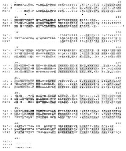
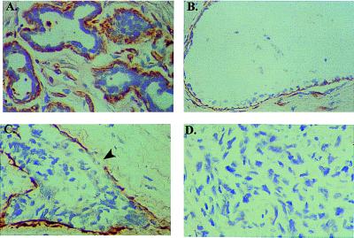
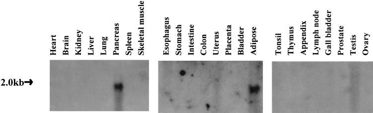
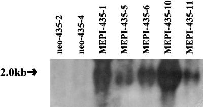
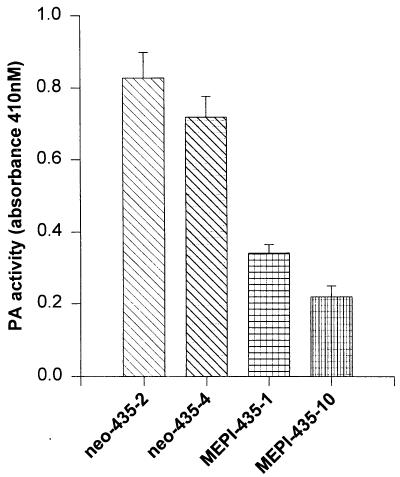
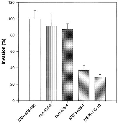
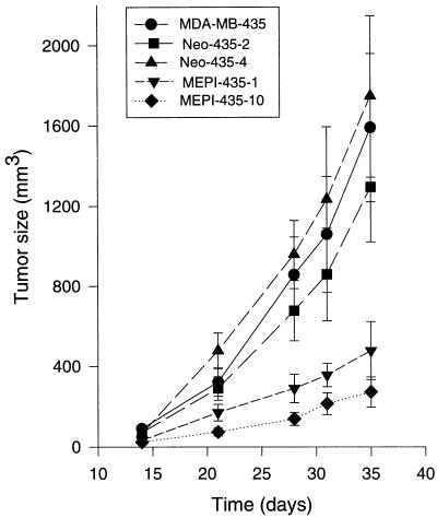
A serpin was identified in normal mammary gland by differential cDNA sequencing. In situ hybridization has detected this serpin exclusively in the myoepithelial cells on the normal and noninvasive mammary epithelial side of the basement membrane and thus was named myoepithelium-derived serine proteinase inhibitor (MEPI). No MEPI expression was detected in the malignant breast carcinomas. MEPI encodes a 405-aa precursor, including an 18-residue secretion signal with a calculated molecular mass of 46 kDa. The predicted sequence of the new protein shares 33% sequence identity and 58% sequence similarity to plasminogen activator inhibitor (PAI)-1 and PAI-2. To determine whether MEPI can modulate the in vivo growth and progression of human breast cancers, we transfected a full-length MEPI cDNA into human breast cancer cells and studied the orthotopic growth of MEPI-transfected vs. control clones in the mammary fat pad of athymic nude mice. Overexpression of MEPI inhibited the invasion of the cells in the in vitro invasion assay. When injected orthotopically into nude mice, the primary tumor volumes, axillary lymph node metastasis, and lung metastasis were significantly inhibited in MEPI-transfected clones as compared with controls. The expression of MEPI in myoepithelial cells may prevent breast cancer malignant progression leading to metastasis.


Figures

Figure 1
Figure 1
Comparison of the predicted amino acid sequence of MEPI with PAI-1 and PAI-2. The available amino acid sequence of PAI-1 and PAI-2 were obtained from the SwissProt database and aligned with the MEPI deduced sequence by using the clustal method of the megalign program from the dnastar software package. Conserved amino acids are shaded. The putative 18 hydrophobic signal peptide is located between two arrows.
Figure 2
Figure 2
In situ hybridization analysis of MEPI expression in human breast. Cells stained brown indicate MEPI gene expression. All sctions were counterstained lightly with hematoxylin for viewing negatively stained epithelial and stromal cells. (A) Myoepithelial cells surrounding lobules from a normal breast-reduction mammoplasty specimen showed strong MEPI expression. (B) A strong positive staining of MEPI in myoepithelial cells surrounding a normal duct. (C) A DCIS showed a partial MEPI expression; arrow indicates the loss of MEPI expression. (D) Negative staining of MEPI in an infiltrating breast cancer. A total of 30 clinical breast specimens were analyzed. Five of five normal breast-reduction mammoplasty samples and five of five benign hyperplasias showed strong expression of MEPI in myoepithelial cells as a continuing layer. Nine of 15 DCIS expressed MEPI in the myoepithelial cells as a continuing layer and the the other 6 DCIS showed partial expression. Five of five infiltrating breast carcinomas were negative. The normal breast section was also hybridized with the sense probe, and no detectable background staining was observed at the same conditions for the antisense probe. All of the sections presented in the figure were derived from the same experiment.
Figure 3
Figure 3
The expression of MEPI gene in a variety of normal human adult tissues. Three blots containing approximately 20 μg of total RNA per line for the above tissues were purchased from Invitrogen. By using a full-length cDNA hybridization probe, a high abundance of 2-kb transcripts was detected in pancreas and adipose tissue.
Figure 4
Figure 4
Northern blot analysis of MEPI transfection of MDA-MB-435 cells. Total RNAs were isolated from two control pCI-neo-transfected clone and five MEPI-transfected clones. Strong MEPI transcripts were detected in MEPI-positive clones. In contrast, no endogenous MEPI transcripts were detected in control clones. The integrity of the RNAs and loading control were ascertained by visualization of the 18S rRNA bands in stained gel (data not shown).
Figure 5
Figure 5
The activities of the CM from MEPI-435 and neo-435 clones on plasminogen activation. CM were collected, concentrated, normalized, and subjected to plasminogen activation analysis as described in Materials and Methods. The numbers represent the mean ± SE of three measurements. Statistical comparisons for pooled MEPI-435 clones relative to pooled neo-435 clones indicated P < 0.001 for the PA activity.
Figure 6
Figure 6
Inhibition of cell invasion by MEPI. Comparison of invasion potentials of MEPI-positive and MEPI-negative cells. The invasion of MDA-MB-435 cells was used as control and was taken as 100%. The invasion potentials of all of the other clones were expressed as a percentage of the control. The numbers represent the mean ± SE of three cultures. Statistical comparisons for pooled MEPI-positive MEPI-435–1 and MEPI-435–10 clones relative to pooled MEPI negative MDA-MB-435, neo-435–2, and neo-435–4 clones indicated P < 0.001 for the invasion.
Figure 7
Figure 7
In vivo tumor growth of MDA-MB-435, neo-435–2, neo-435–4, MEPI-435–1, and MEPI-435–10 cells in nude mice. Each of the eight mice in each group received two injections (one on each side) in the mammary fat pads between the first and second nipples. Tumor size was determined at intervals by three-dimensional measurements (mm) by using a caliper. Only measurable tumors were used to calculate the mean tumor volume for each clone at each time point. Each point represents the mean of tumors ± SE (bars).

References

    1. Andreasen P A, Kjoller L, Duffy M J. Int J Cancer. 1997;72:1–22. - PubMed
    1. Schmitt M, Harbeck N, Thomssen C, Wilhelm O, Magdolen V, Reuning U, Ulm K, Hofler H, Janike F, Graeff H. Thromb Haemostasis. 1997;78:285–296. - PubMed
    1. Duggan C, Kennedy S, Kramer M-D, Barnes C, Elvin P, McDermott E, O’Higgins N, Duffy M J. Br J Cancer. 1997;76:622–627. - PMC - PubMed
    1. Schmitt, M., Janicke, F. & Graeff, F. (1992) Fibrinolysis6, Suppl. 4, 3–26.
    1. Schmitt M, Wilhelm O, Janicke F, Magdolen V, Reuning U, Ohi H, Moniwa N, Kobayashi H, Weidle U, Graeff H. J Obstet Gynaecol Br Emp. 1995;21:151–165. - PubMed

Publication types

MeSH terms

Associated data