. 1999 Mar 30;96(7):3706-11.
doi: 10.1073/pnas.96.7.3706.

GADD45 induction of a G2/M cell cycle checkpoint

Affiliations

GADD45 induction of a G2/M cell cycle checkpoint

X W Wang et al. Proc Natl Acad Sci U S A. .

Abstract

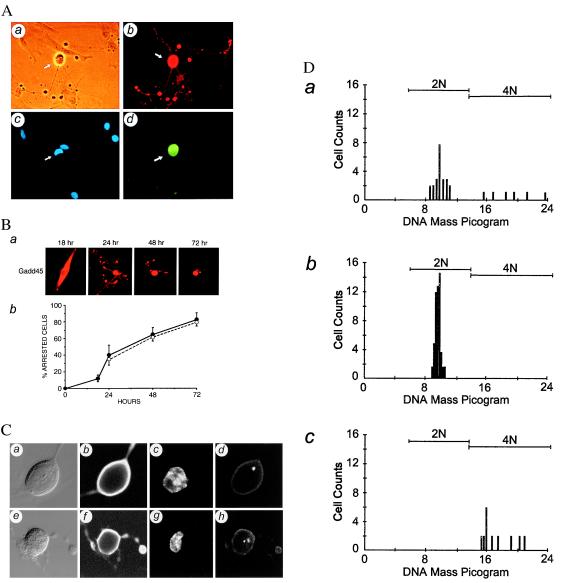
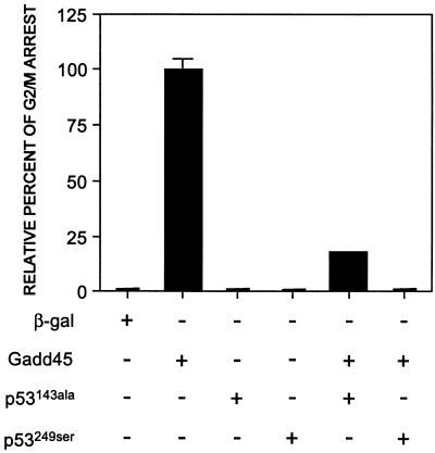
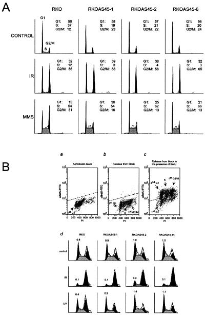
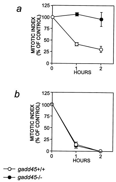
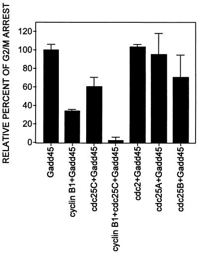
G1/S and G2/M cell cycle checkpoints maintain genomic stability in eukaryotes in response to genotoxic stress. We report here both genetic and functional evidence of a Gadd45-mediated G2/M checkpoint in human and murine cells. Increased expression of Gadd45 via microinjection of an expression vector into primary human fibroblasts arrests the cells at the G2/M boundary with a phenotype of MPM2 immunopositivity, 4n DNA content and, in 15% of the cells, centrosome separation. The Gadd45-mediated G2/M arrest depends on wild-type p53, because no arrest was observed either in p53-null Li-Fraumeni fibroblasts or in normal fibroblasts coexpressed with p53 mutants. Increased expression of cyclin B1 and Cdc25C inhibited the Gadd45-mediated G2/M arrest in human fibroblasts, indicating that the mechanism of Gadd45-mediated G2/M checkpoint is at least in part through modulation of the activity of the G2-specific kinase, cyclin B1/p34(cdc2). Genetic and physiological evidence of a Gadd45-mediated G2/M checkpoint was obtained by using GADD45-deficient human or murine cells. Human cells with endogenous Gadd45 expression reduced by antisense GADD45 expression have an impaired G2/M checkpoint after exposure to either ultraviolet radiation or methyl methanesulfonate but are still able to undergo G2 arrest after ionizing radiation. Lymphocytes from gadd45-knockout mice (gadd45 -/-) also retained a G2/M checkpoint initiated by ionizing radiation and failed to arrest at G2/M after exposure to ultraviolet radiation. Therefore, the mammalian genome is protected by a multiplicity of G2/M checkpoints in response to specific types of DNA damage.


Figures

Figure 1
Figure 1
Gadd45-mediated G2/M cell cycle arrest in normal primary human fibroblasts. (A) At 24 hr, Gadd45-positive cells were identified by double-immunostaining with anti-Gadd45 antibody (b) and a mitosis-specific mAb MPM2 (d). A phase-contrast photomicrograph (a) and a DAPI stain (c) of the same field as b and d. (B) Time course study of Gadd45-mediated G2/M cell cycle arrest was analyzed at 18 hr, 24 hr, 48 hr, or 72 hr. Gadd45-positive cells were counted, and cells displaying a distinct mitosis-like morphology with extensive cytoplasmic extensions were scored as arrested cells. Representative Gadd45-positive cells stained with anti-Gadd45 antibody at the indicated time point (a) and the quantitative data (●) obtained from three independent time course experiments, expressed as mean +/− SD (b), are shown. ○ represent a percentage of the BrdUrd-labeled cells from the same population as an indicator of the ability of these primary cultured human fibroblasts to enter into cell cycle. (C) Gadd45-arrested cells display partially condensed nuclei and contain separated or nonseparated centrosomes, identified by double-immunostaining with anti-Gadd45 antibody (b and f) and a centrosome-specific antiserum (d and h) analyzed by confocal microscopy. The phase-contrast image (a and e) and DAPI-stained nucleus (c and g) of the same cell also are shown. (D) The DNA ploidy of a logarithmically growing fibroblast population (a), cells that were serum-starved for 3 days (b), or cells with Gadd45-induced distinct morphology (c) also were analyzed by Feulgen staining and CAS image analysis.
Figure 2
Figure 2
Modulation of Gadd45-induced G2/M arrest by p53 mutants in normal primary human fibroblasts. Cells were either microinjected alone with β-gal, GADD45, and two p53 mutants or coinjected with GADD45 and the p53 mutants. Gadd45-positive cells displaying an arrested morphology were scored at 24 hr. Anti-p53 CM-1 polyclonal antibody was used to screen for p53-positive cells. At least three independent microinjection experiments were averaged, and data are expressed as the mean ± SD.
Figure 3
Figure 3
Defect in a UV-induced G2/M checkpoint in Gadd45-deficient cells. (A) Flow cytometric analysis of the human colon carcinoma cell line RKO and its GADD45 antisense overexpressing clones in response to IR or MMS treatment. Unsynchronized cells were treated with either 6.3 Gy IR or 25 μg/ml MMS and analyzed by FACScan after a 24-hr incubation. The G1, S, and G2/M fractions are indicated, as calculated by modfit analysis. (B) Increased expression of antisense GADD45 allows RKO cells to escape the G2/M checkpoint arrest induced by UV-induced damage. Parental RKO cells and three antisense GADD45-expressing clones were synchronized with aphidicolin for 24 hr (>95% at G1) and release from blocking in the presence of BrdUrd. (ac) Representative profiles of RKO cells subjected to synchronization. The intensities of fluorescein isothiocyanate (anti-BrdUrd signal) and PI (DNA contents) are indicated. Three antisense GADD45 clones show similar synchronization profiles as their parental RKO cells (data not shown). On release from late G1, cells were treated with 6.3 Gy IR or 5 J/m2 UV and incubated for an additional 20 hr in the presence of BrdUrd. The BrdUrd-positive cells were subjected to FACScan and modfit analysis (d). The first solid peaks, the gray area, and the second solid peaks represent the second G1 (2nd G1), first S (S) and first G2/M (1st G2) fractions, respectively. No second S phase should be observed in this condition. The numbers above the second G1 peaks are the ratio of G1/G2 (percent of G1 fraction escaped from G2/M arrest).
Figure 4
Figure 4
Failure of the UV-induced mitotic delay of murine gadd45 (−/−) lymphocytes. Proliferating lymphocytes from gadd45 (+/+) mice (○) or gadd45 (−/−) mice (●) were treated with UV radiation at 7.5 J/m2 (A) or with IR at 5 Gy (B). Mitotic indices were determined at the indicated times after irradiation and are shown as a percentage of the appropriate unirradiated control cells. For A, pooled lymphocytes from two gadd45 (−/−) male mice at 11 weeks of age and two gadd45 (+/+) male mice at 15 weeks of age were used. For B, pooled lymphocytes from three gadd45 (−/−) male mice at 4 weeks of age and three gadd45 (+/+) male mice at 6 weeks of age were used. Similar results were observed from three separate cultures that also included female mice (not shown).
Figure 5
Figure 5
Effect of various cell cycle-modulating genes on the Gadd45-induced G2/M arrest. Normal primary human fibroblasts were either microinjected alone with GADD45 or coinjected with cyclin B1, cdc25A, cdc25B, cdc25C, or p34cdc2 (cdc2). Cells were fixed at 24 hr, and Gadd45-positive cells displaying an arrested morphology were scored. At least three independent microinjection experiments were averaged, and data are expressed as mean ± SD. By using Student’s t test, a statistically significant P value (<0.05) was obtained between GADD45 alone and coinjection with cyclin B1 and/or cdc25C.

References

    1. Hartwell L H, Weinert T A. Science. 1989;246:629–634. - PubMed
    1. Cox L S, Lane D P. BioEssays. 1995;17:501–508. - PubMed
    1. Gottlieb T M, Oren M. Biochim Biophys Acta. 1996;1287:77–102. - PubMed
    1. Ko L J, Prives C. Genes Dev. 1996;10:1054–1072. - PubMed
    1. Levine A J. Cell. 1997;88:323–331. - PubMed

MeSH terms