. 1999 Mar 30;96(7):3757-62.
doi: 10.1073/pnas.96.7.3757.

Probing the Saccharomyces cerevisiae centromeric DNA (CEN DNA)-binding factor 3 (CBF3) kinetochore complex by using atomic force microscopy

Affiliations

Probing the Saccharomyces cerevisiae centromeric DNA (CEN DNA)-binding factor 3 (CBF3) kinetochore complex by using atomic force microscopy

L I Pietrasanta et al. Proc Natl Acad Sci U S A. .

Abstract

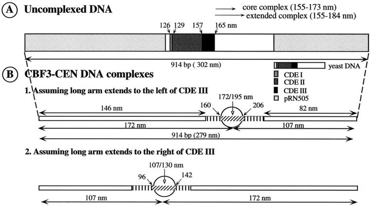
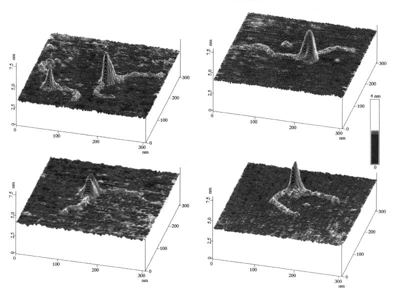
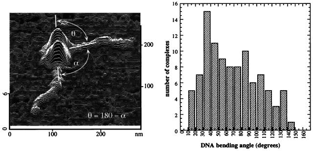
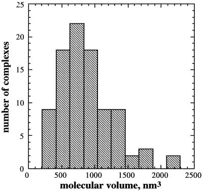
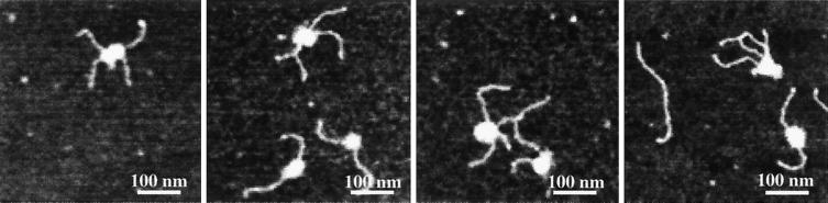
Yeast centromeric DNA (CEN DNA) binding factor 3 (CBF3) is a multisubunit protein complex that binds to the essential CDEIII element in CEN DNA. The four CBF3 proteins are required for accurate chromosome segregation and are considered to be core components of the yeast kinetochore. We have examined the structure of the CBF3-CEN DNA complex by atomic force microscopy. Assembly of CBF3-CEN DNA complexes was performed by combining purified CBF3 proteins with a DNA fragment that includes the CEN region from yeast chromosome III. Atomic force microscopy images showed DNA molecules with attached globular bodies. The contour length of the DNA containing the complex is approximately 9% shorter than the DNA alone, suggesting some winding of DNA within the complex. The measured location of the single binding site indicates that the complex is located asymmetrically to the right of CDEIII extending away from CDEI and CDEII, which is consistent with previous data. The CEN DNA is bent approximately 55 degrees at the site of complex formation. A significant fraction of the complexes are linked in pairs, showing three to four DNA arms, with molecular volumes approximately three times the mean volumes of two-armed complexes. These multi-armed complexes indicate that CBF3 can bind two DNA molecules together in vitro and, thus, may be involved in holding together chromatid pairs during mitosis.


Figures

Figure 1
Figure 1
Location of the CBF3 complex on yeast CEN DNA. (A) Scaled diagram of the linear 914-bp CEN DNA NdeI/AflIII restriction fragment showing the locations (in nanometers, numbered from the left) of CDEI, CDEII, and CDEIII. (B) Scaled diagrams illustrate this DNA molecule containing a CBF3 complex. The region of DNA covered by the CBF3 complex is indicated by a circle and the 23-nm reduction in contour length is indicated by a vertical arrow within the circle. The diameter of the circle shows the width of the complex at half-maximal height. The vertical stripes on each side of the circle indicate the apparent width of the base of the CBF3 complex, which is broadened by an amount comparable to the width of the AFM tip. In diagram B-1 the location of the complex relative to CDEI and CDEIII assumes that the long DNA arm extends to the left of CDEIII. In B-2, it is assumed that the long DNA arm extends to the right of CDEIII. All measurements are in nm. The core and extended complexes are indicated according to refs. and .
Figure 2
Figure 2
AFM images of CBF3–CEN DNA complexes on mica. The images are presented as line plots at a 30° viewing angle to emphasize topography. Height is indicated by the gray-scale code bar.
Figure 3
Figure 3
CEN DNA is bent at the site of CBF3 binding. (Left) A typical CBF3–CEN DNA complex showing the measured angle α and its relation to the bend angle θ. (Right) Histogram of the DNA bend angles measured from AFM images of 94 CBF3–CEN DNA complexes.
Figure 4
Figure 4
Molecular volumes of CBF3–CEN DNA complexes. Histogram of the calculated volumes of the CBF3–CEN DNA complexes using the bearing volume method to analyze the AFM images (see Materials and Methods).
Figure 5
Figure 5
Multi-armed CBF3–CEN DNA complexes. AFM images (top views) of complexes containing single and multiple DNA molecules. (Image sizes are 500 × 500 nm.)

References

    1. Lechner J, Ortiz J. FEBS Lett. 1996;389:70–74. - PubMed
    1. Lechner J, Carbon J. Cell. 1991;64:717–725. - PubMed
    1. Espelin C W, Kaplan K B, Sorger P K. J Cell Biol. 1997;139:1383–1396. - PMC - PubMed
    1. Bustamante C, Rivetti C. Annu Rev Biophys Biomol Struct. 1996;25:395–429. - PubMed
    1. Bustamante C, Rivetti C, Keller D J. Curr Opin Struct Biol. 1997;7:709–716. - PubMed

Publication types

MeSH terms