. 1999 Mar 30;96(7):3819-23.
doi: 10.1073/pnas.96.7.3819.

Pathogenetic sequence for aneurysm revealed in mice underexpressing fibrillin-1

Affiliations

Pathogenetic sequence for aneurysm revealed in mice underexpressing fibrillin-1

L Pereira et al. Proc Natl Acad Sci U S A. .

Abstract

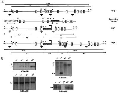
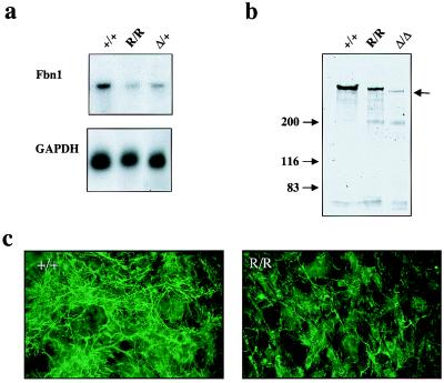
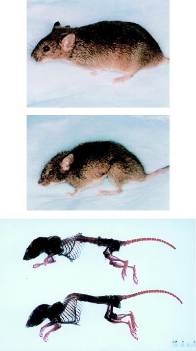
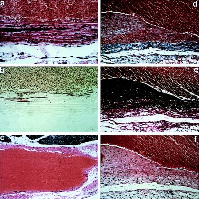
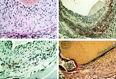
Dissecting aortic aneurysm is the hallmark of Marfan syndrome (MFS) and the result of mutations in fibrillin-1, the major constituent of elastin-associated extracellular microfibrils. It is yet to be established whether dysfunction of fibrillin-1 perturbs the ability of the elastic vessel wall to sustain hemodynamic stress by disrupting microfibrillar assembly, by impairing the homeostasis of established elastic fibers, or by a combination of both mechanisms. The pathogenic sequence responsible for the mechanical collapse of the elastic lamellae in the aortic wall is also unknown. Targeted mutation of the mouse fibrillin-1 gene has recently suggested that deficiency of fibrillin-1 reduces tissue homeostasis rather than elastic fiber formation. Here we describe another gene-targeting mutation, mgR, which shows that underexpression of fibrillin-1 similarly leads to MFS-like manifestations. Histopathological analysis of mgR/mgR specimens implicates medial calcification, the inflammatory-fibroproliferative response, and inflammation-mediated elastolysis in the natural history of dissecting aneurysm. More generally, the phenotypic severity associated with various combinations of normal and mutant fibrillin-1 alleles suggests a threshold phenomenon for the functional collapse of the vessel wall that is based on the level and the integrity of microfibrils.


Figures

Figure 1
Figure 1
Fbn1 gene targeting. (a) From top to bottom: the wild-type region that was targeted with the position of the probes (A–D) and the restriction sites used in the Southern analysis; the targeting vector with the arrows indicating the direction of transcription of the selectable marker genes; and the mgΔ and mgR alleles with the BamHI and HindIII fragments. (b) Southern blots of tail DNA from mice with different Fbn1 genotypes digested with the indicated enzymes and hybridized to the indicated probes. The samples include wild-type (+/+), heterozygous (Δ/+), and homozygous (Δ/Δ) mgΔ mice and heterozygous (R/+) and homozygous (R/R) mgR mice.
Figure 2
Figure 2
Characterization of the mgR products. (a) Northern blot hybridization of poly(A)+ RNA purified from pooled skin samples hybridized to Fbn1 and GAPDH probes. The normalized Fbn1 mRNA expression levels for mgR/mgR and mgΔ/+ tissues, relative to +/+ controls, were 0.28 and 0.52, respectively. (b) SDS/PAGE of immunoprecipitated metabolically labeled fibrillin-1 from the medium of cells established from wild-type and mutant animals; arrow points to the fibrillin-1 products. (c) Immunofluorescence of cultured dermal fibroblasts from mgR/mgR (R/R) and wild-type (+/+) littermates by fibrillin-1 antisera; cells were processed 72 hr after plating.
Figure 3
Figure 3
Clinical presentation of wild-type (Top) and homozygous mgR (Middle) littermates demonstrating severe kyphosis and overgrowth of the ribs. Alizarin red and alcian blue staining of bone and cartilage, respectively, after removal of soft tissues (Bottom).
Figure 4
Figure 4
Histopathology of homozygous mgR mice examined between 6 wk and 3 mo of postnatal life. (a and b) Calcification of elastic lamellae in the aortic media at 6 wk; staining with hemotoxylin and eosin (a) or alizarin red (b). (c) Aneurysmal dilatation with vessel-wall thinning and calcification of elastic lamellae at 3 mo; hemotoxylin and eosin. (×15) (d–f) Intimal hyperplasia with accumulation of excessive collagen and elastin and smooth muscle-cell proliferation and monocyctic infiltration with fragmentation of elastic lamellae in the media at 3 mo. Staining was performed with trichrome blue (d), Verhoeff–van Gieson stain (e), or hemotoxylin and eosin (f). The vessel lumen is at top except in c. [×250 (a, b, d–f).]
Figure 5
Figure 5
Histopathology and immunohistochemistry of homozygous mgR mice examined between 3 and 6 mo of postnatal life. (a) Intimal hyperplasia and disorganized elastic lamellae at 3 mo; hemotoxylin and eosin. (b and c) Horseradish peroxidase immunostaining using mAb F4/80 specific for mature macrophages performed on 3-mo (b) and 5–1/2-mo (c) specimens. (c) Extensive macrophage infiltration at the adventitial border associated with aneurysmal dilatation. (d) Bony metaplasia with hematopoiesis within the vessel wall at the junction of the aortic annulus and the ascending aorta at 6 mo; Verhoeff–van Gieson. (×350.)
Figure 6
Figure 6
SDS/PAGE analysis of MMP-12 cleavage of fibrillin-1. Immunoprecipitated metabolically labeled fibrillin was incubated without (lane 2) or with (lane 3) recombinant MMP-12; in lane 1, radioactive fibronectin eluted from gelatin–Sepharose was included as a marker. The ≈200-kDa band seen in lane 2 is an MMP-12-sensitive contaminant of the immunoprecipitation.

References

    1. Mecham R P, Davies E. In: Extracellular Matrix Assembly and Structure. Yurchenco P D, Birk D E, Mecham R P, editors. New York: Academic; 1994. pp. 281–314.
    1. Dietz H, Ramirez F, Sakai L. In: Advances in Human Genetics. Harris H, Hirschhorn K, editors. Vol. 22. New York: Plenum; 1994. pp. 153–186. - PubMed
    1. Dietz H, Pyeritz R. Hum Mol Genet. 1995;4:1799–1809. - PubMed
    1. Ramirez F. Curr Opin Genet Dev. 1996;6:309–315. - PubMed
    1. Pereira L, Andrikopoulos K, Tian J, Lee S Y, Keene D R, Ono R, Reinhardt D P, Sakai LY, Jensen-Biery N, Bunton T, et al. Nat Genet. 1997;17:218–222. - PubMed

Publication types

MeSH terms