. 1999 Mar 30;96(7):3888-93.
doi: 10.1073/pnas.96.7.3888.

The Ets transcription factor ERM is Th1-specific and induced by IL-12 through a Stat4-dependent pathway

Affiliations

The Ets transcription factor ERM is Th1-specific and induced by IL-12 through a Stat4-dependent pathway

W Ouyang et al. Proc Natl Acad Sci U S A. .

Abstract

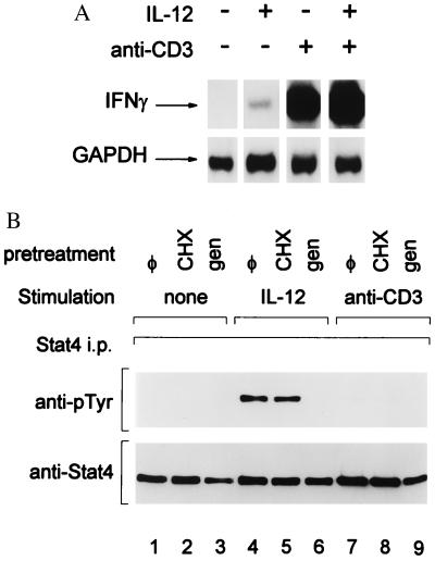
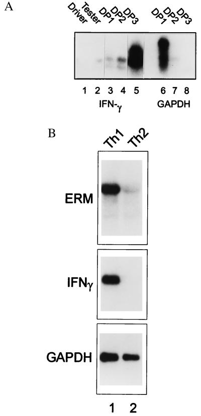
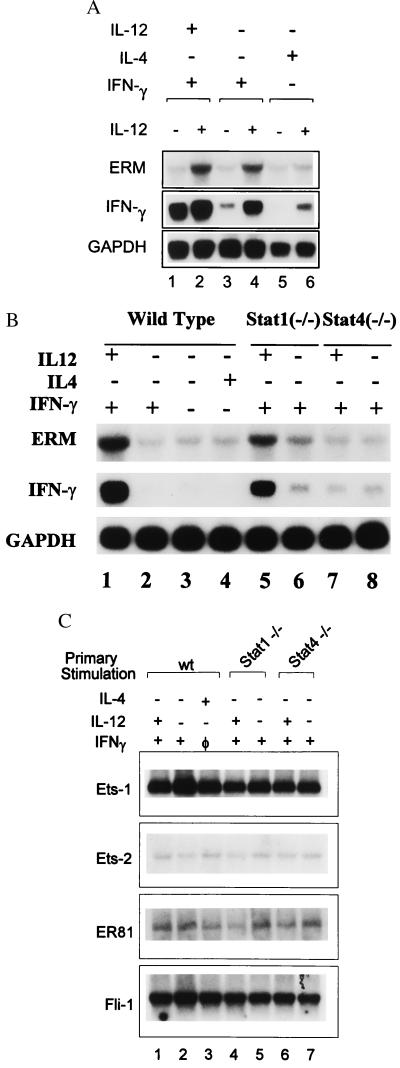
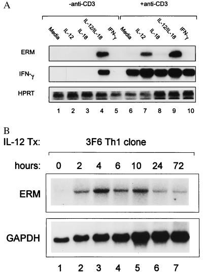
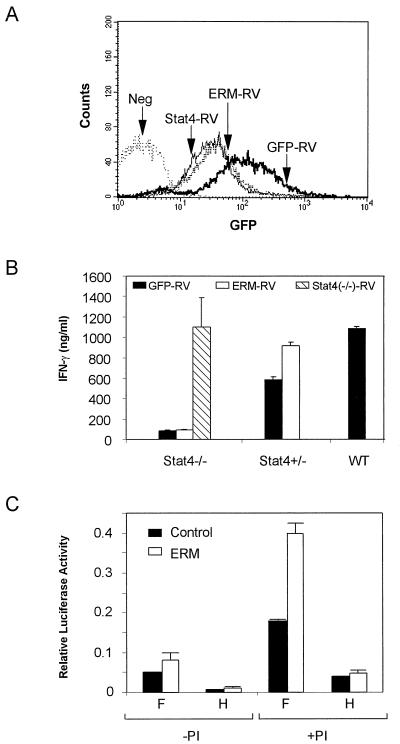
Interleukin 12 (IL-12)-induced T helper 1 (Th1) development requires Stat4 activation. However, antigen-activated Th1 cells can produce interferon gamma (IFN-gamma) independently of IL-12 and Stat4 activation. Thus, in differentiated Th1 cells, factors regulated by IL-12 and Stat4 may be involved in IFN-gamma production. Using subtractive cloning, we identified ERM, an Ets transcription factor, to be a Th1-specific, IL-12-induced gene. IL-12-induction of ERM occurred in wild-type and Stat1-deficient, but not Stat4-deficient, T cells, suggesting ERM is Stat4-inducible. Retroviral expression of ERM did not restore IFN-gamma production in Stat4-deficient T cells, but augmented IFN-gamma expression in Stat4-heterozygous T cells. Ets factors frequently regulate transcription via cooperative interactions with other transcription factors, and ERM has been reported to cooperate with c-Jun. However, in the absence of other transcription factors, ERM augmented expression of an IFN-gamma reporter by only 2-fold. Thus, determining the requirement for ERM in Th1 development likely will require gene targeting.


Figures

Figure 1
Figure 1
IFN-γ production induced by anti-CD3 stimulation in differentiated Th1 cells is IL-12- and Stat4-indepenent. (A) 3F6 T cells were stimulated (+) with anti-CD3 or IL-12 (10 units/ml) as indicated or left unstimulated (−) for 4 hr as described in Methods. Total RNA was prepared and Northern blot analysis for IFN-γ was performed (17). The autoradiogram was overexposed to allow detection of the faint band present in the IL-12-treated lane. The blot was stripped and reprobed for GAPDH as a loading control as described (17). (B) 3F6 T cells were pretreated with Genistein (50 μg/ml) (gen) or cycloheximide (10 μg/ml) (CHX) or left untreated (φ), as indicated, for 30 min and then stimulated with IL-12 (10 units/ml) or anti-CD3 for 30 min, or untreated (none) as indicated. Cells were lysed, and immunoprecipitation for Stat4 was performed by using NB34 as described (17). Western detection of phosphotyrosine was performed with antiphosphotyrosine RC20 (Transduction Laboratories), and blots were stripped and reprobed with anti-Stat4 (Lower) to indicate uniform Stat4 immunoprecipitation.
Figure 2
Figure 2
Cloning of Th1-specific genes by RDA. (A) Two hundred-microgram PCR products of Driver (Th1) and Tester (Th2), as well as each difference subtraction product DP1, DP2, and DP3, were electrophoresed on 1.5% agarose gel and transferred to Zeta membrane. The membrane was probed by either IFN-γ or GAPDH cDNA probe as indicated. (B) Splenocytes from DO11.10 TCR-transgenic mice were activated with OVA and APCs with either IL-12 or IL-4, as indicated, to initiate Th1 development (lane 1) or Th2 development (lane 2) for 48 hr. Total RNA was prepared and Northern blot analysis was performed by using a full-length ERM cDNA as probe (ERM). The blot was stripped and reprobed for IFN-γ and GAPDH as indicated.
Figure 3
Figure 3
ERM expression is IL-12-inducible and Stat4-dependent. (A) Wild-type DO11.10 TCR-transgenic splenocytes were activated with OVA and APCs plus various combinations of IL-12, IL-4, and IFN-γ as indicated. On day 7, T cells were harvested and restimulated with OVA and APCs, with (+) or without (−) the addition of IL-12 (10 units/ml). Total RNA was isolated after 48 hr, and Northern blot analysis for ERM, IFN-γ, and GAPDH was performed as above. (B) DO11.10 splenocytes from wild-type, Stat1-deficient, or Stat4-deficient mice DO11.10 mice were activated by OVA in the presence (+) or absence (−) of indicated cytokines for 48 hr. RNA was prepared and Northern blot analysis of ERM, IFN-γ, and GAPDH was performed as described in A. (C) The indicated Ets family members were obtained as expressed sequence tag clones from Genome Systems (St. Louis), and their identity was confirmed by DNA sequencing. Probes were derived from unique coding or 3′ untranslated regions.
Figure 4
Figure 4
Regulation of ERM expression by IL-12 and IL-18. (A) Th1 cells were generated from DO11.10 TCR-transgenic splenocytes as described above, restimulated with OVA and APCs, and, 7 days later, restimulated with the indicated combinations of anti-CD3, IL-12 (10 units/ml), IL-18 (50 ng/ml), and IFN-γ (100 units/ml) for 48 hr. Total RNA was prepared and Northern blot analysis of ERM, IFN-γ, and GAPDH was performed. (B) Resting 3F6 T cells were stimulated by using anti-CD3-coated plates and IL-12 (10 units/ml) for the indicated times, total RNA was prepared, and ERM and GAPDH Northern blot analysis was performed as described above.
Figure 5
Figure 5
Effects of ERM expression on IFN-γ production in Stat4-deficient and Stat4-herterozygous T cells. (A) Stat4-deficient, Stat4-heterozygous, and wild-type DO11.10 TCR-transgenic T cells were activated in vitro by using OVA/APCs and infected on day 1 after primary activation with either empty vector (GFP-RV), Stat4-expresing retrovirus (Stat4-RV), or ERM-expressing retrovirus (ERM-RV). Infected cells were purified by cell sorting on day 7 for GFP (FL1) and CD4 expression by using anti-mouse-CD4-Phycoerythrin (PharMingen). Sorted GFP+/CD4+ T cells were reactivated with OVA, APCs, and IL-12 (see Methods), and stable transfection was confirmed by GFP expression by FACS analysis 3 days later. Data shown are single color histograms of the indicated sorted transfectant populations. The negative control (Neg) is an uninfected, Stat4-deficient DO11.10 T cell population activated concurrently under the same conditions. (B) Retrovirally infected T cells from the indicated populations described in A were harvested, washed, and restimulated at 1.25 × 10⁵/ml with 0.3 μM of OVA and irradiated BALB/c splenocytes as APCs. Supernatants were harvested after 48 hr, and IFN-γ production was determined by ELISA. Similar results were obtained in three similar independent experiments. (C) Luciferase reporter assays were performed as described in Methods by using the IFN-γ promoter constructs F and H. Transfected cells were treated with phorbol 12-myristate 13-acetate and ionomycin (PI+) or left untreated (PI−) as described (25). Data are presented as relative light units after normalization for transfection efficiency by using CMV-Renilla luciferase.

References

    1. Abbas A K, Murphy K M, Sher A. Nature (London) 1996;383:787–793. - PubMed
    1. Seder R A, Paul W E. Annu Rev Immunol. 1994;12:635–673. - PubMed
    1. Hsieh C S, Macatonia S E, O’Garra A, Murphy K M. Int Immunol. 1993;5:371–382. - PubMed
    1. Macatonia S E, Hosken N A, Litton M, Vieira P, Hsieh C S, Culpepper J A, Wysocka M, Trinchieri G, Murphy K M, O’Garra A. J Immunol. 1995;154:5071–5079. - PubMed
    1. Manetti R, Parronchi P, Giudizi M G, Piccinni M-P, Maggi E, Trinchieri G, Romagnani S. J Exp Med. 1993;177:1199–1204. - PMC - PubMed

Publication types

MeSH terms