. 1999 Mar 30;96(7):3906-10.
doi: 10.1073/pnas.96.7.3906.

Sustained correction of bleeding disorder in hemophilia B mice by gene therapy

Affiliations

Sustained correction of bleeding disorder in hemophilia B mice by gene therapy

L Wang et al. Proc Natl Acad Sci U S A. .

Abstract

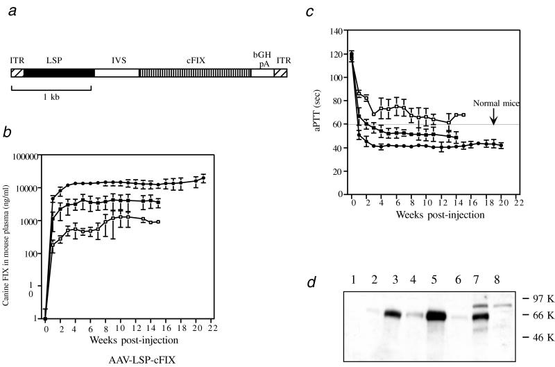
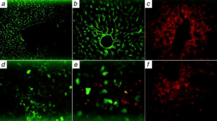
Mice generated by disrupting the clotting factor IX gene exhibit severe bleeding disorder and closely resemble the phenotype seen in hemophilia B patients. Here we demonstrate that a single intraportal injection of a recombinant adeno-associated virus (AAV) vector encoding canine factor IX cDNA under the control of a liver-specific enhancer/promoter leads to a long-term and complete correction of the bleeding disorder. High level expression of up to 15-20 microgram/ml of canine factor IX was detected in the plasma of mice injected with 5.6 x 10(11) particles of an AAV vector for >5 months. The activated partial thromboplastin time of the treated mice was fully corrected to higher than normal levels. Liver-specific expression of canine factor IX was confirmed by immunofluorescence staining, and secreted factor IX protein was identified in the mouse plasma by Western blotting. All treated mice survived the tail clip test without difficulty. Thus, a single intraportal injection of a recombinant adeno-associated virus vector expressing factor IX successfully cured the bleeding disorder of hemophilia B mice, proving the feasibility of using AAV-based vectors for liver-targeted gene therapy of genetic diseases.


Figures

Figure 1
Figure 1
Long-term expression of functional canine factor IX in hemophilia B mice. (a) Schematic drawing of rAAV-LSP-cFIX vector. (b) ELISA assays. Recombinant AAV-LSP-cFIX was delivered as a single intraportal injection into the liver of adult hemophilic C57BL/6 mice in a dose of 2 × 10¹¹ (n = 4, ■) or 5.6 × 10¹¹ (n = 5, ●) vector genomes or into the liver of adult hemophilic 129 mice at a dose of 2 × 10¹¹ particles (n = 3, □). Mice were bled weekly after vector administration. The canine factor IX concentration in the mouse plasma was determined by an ELISA assay. (c) Functional factor IX activity in the mouse plasma was determined by an in vitro aPTT assay (same symbols as in b). The line in the middle shows the aPTT for normal mice. (d) Western blot analysis. Plasma from C57BL/6 hemophilic mice 4 months postinjection with 2.8 × 10¹¹ particles or 5.6 × 10¹¹ particles of AAV-LSP-cFIX was analyzed by Western blotting. Lanes: 1, naive mouse, (0.1 μl of plasma); 2 and 3, plasma from mouse injected with 2.8 × 10¹¹ particles (0.01 and 0.1 μl, respectively); 4 and 5, plasma from mouse injected with 5.6 × 10¹¹ particles (0.01 and 0.1 μl, respectively); 6 and 7, normal canine plasma (0.01 and 0.1 μl, respectively) (Sigma); 8, 0.1 μl of plasma from a hemophilic dog (a gift from T. Nichols of the University of North Carolina, Chapel Hill).
Figure 2
Figure 2
Detection of canine factor IX in the mouse liver by immunofluorescence staining. AAV-LSP-cFIX (2.8 × 10¹¹ particles in 200 μl) was injected intraportally into hemophilic C56BL/6 mice. Four months after the injection, one mouse was killed, and the liver was analyzed for canine factor IX by immunofluorescence staining. Plasma level of canine factor IX was 6.5 μg/ml at the time the mouse was killed. Shown is AAV-transduced liver demonstrating the secreted canine factor IX in the linings of sinusoids and central vein (a, ×20; b, ×40). (c) Control naive mouse (×40). (df) Transduction of mouse liver by AAV-LSP-GFP. Particles (3 × 10¹¹) in 150 μl were injected intraportally into C56BL/6 mice. Eighteen weeks after the injection, one mouse was killed, and the liver was analyzed for GFP expression by immunofluorescence microscopy. Hepatocytes positive for GFP expression were identified in the AAV-transduced liver. [d, ×20; e, ×40; f (control naive mouse), ×40].

Similar articles

Cited by

References

    1. Lin H F, Maeda N, Smithies O, Straight D L, Stafford D W. Blood. 1997;90:3962–3966. - PubMed
    1. Wang L, Zoppe M, Hackeng T M, Griffin J H, Lee K-F, Verma I M. Proc Natl Acad Sci USA. 1997;94:11563–11566. - PMC - PubMed
    1. Kundu R K, Sangiorgi F, Wu L Y, Kurachi K, Anderson W F, Maxson R, Gordon E M. Blood. 1998;92:168–174. - PubMed
    1. Kung S H, Hagstrom J N, Cass D, Tai S J, Lin H F, Stafford D W, High K A. Blood. 1998;91:784–790. - PubMed
    1. Xiao X, Li J, Samulski R J. J Virol. 1996;70:8098–8108. - PMC - PubMed

Publication types

MeSH terms