. 1999 Mar 30;96(7):3916-21.
doi: 10.1073/pnas.96.7.3916.

RB-mediated suppression of spontaneous multiple neuroendocrine neoplasia and lung metastases in Rb+/- mice

Affiliations

RB-mediated suppression of spontaneous multiple neuroendocrine neoplasia and lung metastases in Rb+/- mice

A Y Nikitin et al. Proc Natl Acad Sci U S A. .

Abstract

Alterations in pathways mediated by retinoblastoma susceptibility gene (RB) product are among the most common in human cancer. Mice with a single copy of the Rb gene are shown to develop a syndrome of multiple neuroendocrine neoplasia. The earliest Rb-deficient atypical cells were identified in the intermediate and anterior lobes of the pituitary, the thyroid and parathyroid glands, and the adrenal medulla within the first 3 months of postnatal development. These cells form gross tumors with various degrees of malignancy by postnatal day 350. By age of 380 days, 84% of Rb+/- mice exhibited lung metastases from C-cell thyroid carcinomas. Expression of a human RB transgene in the Rb+/- mice suppressed carcinogenesis in all tissues studied. Of particular clinical relevance, the frequency of lung metastases also was reduced to 12% in Rb+/- mice by repeated i.v. administration of lipid-entrapped, polycation-condensed RB complementary DNA. Thus, in spite of long latency periods during which secondary alterations can accumulate, the initial loss of Rb function remains essential for tumor progression in multiple types of neuroendocrine cells. Restoration of RB function in humans may prove an effective general approach to the treatment of RB-deficient disseminated tumors.


Figures
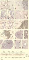
Figure 1
Figure 1
Multiple neuroendocrine neoplasia in Rb+/− mice. (A and B) Gross compound pituitary tumor on P394. The tumor consists of two histologically distinct components that substitute for the pituitary anterior (AL) and intermediate (IL) lobes, which are demarcated by remnants of Rathke’s cleft (arrows). AL tumor cells contain α-GSU (A) and are positioned loosely around sinusoid-like vessels. IL tumor cells contain α-melanocyte-stimulating hormone (B) and form poorly vascularized epithelioid fields with central necrotic and hemorrhagic areas. (C and D) EAP in the anterior pituitary lobe on P90 before (C) and after (D) microdissection for genotype analysis by the PCR. (E) C-cell carcinoma on P340 showing the typical arrangement of polyhedral tumor cells in solid nests and rough hyalinized collagen with calcification (arrow). (F and G) C-cell EAP (arrow) on P60 before (F) and after (G) microdissection. The atypical cells show a parafollicular location. (H) Medullary thyroid carcinoma invading surrounding tissues on P469. Accumulation of calcitonin (brown color) is evident in the cytoplasm of tumor cells (arrow). CR, cartilage. (I) Calcitonin-containing metastatic cells in the lung on P463. The metastatic cells exhibit intraalveolar spreading (arrows). (J) A well-vascularized parathyroid tumor (PG) and a neighboring solid C-cell tumor of the thyroid gland (TG) on P370. (K) Parathyroid hormone expression in parathyroid tumor cells. (L) Pheochromocytoma of the adrenal medulla (AM) compressing the adrenal cortex (AC). (M and N) EAP in the adrenal medulla (AM) on P60 before (M) and after (N) microdissection. The arrow indicates multiple apoptotic figures. Staining: avidin-biotin-peroxidase immunostaining for α-GSU (A), α-melanocyte-stimulating hormone (B), calcitonin (H and I), or parathyroid hormone (K) with hematoxylin counterstaining; (CG, J, and LN) hematoxylin-eosin staining. [Bar: 160 μm (A and B), 40 μm (C, D, and I), 110 μm (E), 60 μm (F and G), 50 μm (H), 150 μm (J), and 20 μm (K), 390 μm (L), and 70 μm (M and N).] (O) Absence of the wild-type Rb allele (151-bp PCR product) in gross tumors (T; lanes 3, 4, 7, 10, 11, 17, and 18) and EAPs (E; lanes 1, 2, 6, 8, 9, 14, and 16) of the pituitary anterior lobe (AL, lanes 1–4), the parathyroid gland (PG, lanes 6 and 7), thyroid C cells (TG, lanes 8–11), lung metastases (L, lanes 12 and 13), or adrenal medulla (lanes 14 and 16–18). N, Rb+/− normal tissue (lanes 5 and 15). Nondenaturing 12% polyacrylamide gel stained with silver. The 236-bp band corresponds to the mutant Rb allele (11).
Figure 2
Figure 2
Characterization of RB transgene expression. (AF) Tetracycline-regulated expression of RB in Rb+/−RTgRB mice. Transgenic littermates were treated with tetracycline hydrochloride (1 mg/ml) from fertilization until either P94 (A, C, and E) or P90 (B, D, and F). On P94 expression of the RB transgene was tested in atypical cells of the pituitary intermediate lobe (A and B), the thyroid gland (C and D), and the adrenal medulla (E and F). In mice continuously exposed to tetracycline no RB is detected in nuclei of atypical (arrows) melanotrophs (A), C cells (C), and medulla cells (E). Withdrawal of tetracycline on P90 results in expression of RB transgene in those cells by P94 (B, D, and F). Note that staining of normal cells reflects expression of either endogenous Rb (A, C, and E, arrowheads) or both endogenous Rb and exogenous RB (B, D, and F). (GK) RB expression in metastatic C cells after administration of LPDs. Mice were killed 24 hr after i.v. injection with either LPD-RB (G, H, and J), LPD-RBH209 (K), or LPD-lacZ (I). (G and H) Groups of metastatic cells before (G) and after (H) microdissection. (IK) Simultaneous detection of cytoplasmic calcitonin and nuclear RB in metastatic cells. Unlike metastatic cells from mice treated with LPD-lacZ (I), those from mice treated with either LPD-RB (J) or LPD-RBH209 (K) showed intense RB staining. Avidin-biotin-peroxidase immunostaining with C-15 antiserum alone (AF) or together with calcitonin-specific antibodies (IK). Counterstaining with methyl green. (G and H) Hematoxylin-eosin staining. [Bar: 40 μm (A and B), 20 μm (CF), 55 μm (G and H), and 16 μm (IK).]
Figure 3
Figure 3
Detection of RB expression after LPD-RB administration. (A) Detection by microdissection and PCR analysis of the RB transgene in metastatic cells from lung tissue of mice treated with either LPD-RB (lanes 2–5) or LPD-RBH209 (H209, lanes 6 and 7) but not in those from mice exposed to LPD-Luc (lane 1). The 114-bp and 194-bp PCR fragments are diagnostic for RB cDNA and the Rb gene, respectively (20). Lane 6, DNA size markers (Amresco, Euclid, OH). (B) Immunoprecipitation and immunoblot analysis of RB expression in lungs of mice treated with LPD-RB (lanes 1–4), LPD-RBH209 (lanes 5 and 6), or in those of mice exposed to LPD-Luc (lanes 7 and 8). Immunoprecipitation (IP) was performed with C-15 antiserum, which recognizes both human RB (110 kDa) and mouse Rb (105 kDa), or mAb 11D7, which is specific for human RB protein, and immunoblot analysis was performed with mAB 245 (Upper). Detection of p84, a nuclear matrix protein (57), was assessed as a control for gel loading (Lower). (C) Detection of Rb and RB (Upper, 161 bp) and β-actin (Lower, 306 bp) mRNAs in the lung (lane 1), thyroid C cells (lane 2), and lung metastases treated with LPD-RB (lane 3), LPD-RBH209 (lane 6), LPD-Luc (lane 4), or LPD-lacZ (lane 7). Lanes 5 and 8, reverse transcriptase (RT)–PCRs without RT. (A and C) Polyacrylamide gel stained with silver. (D) Relative amounts of Rb cDNA (mean ± SD) in the lung (n = 8), C cells (CC, n = 5), and metastatic cells treated with either LPD-RB (RB, n = 6) or LPD-RBH209 (H209, n = 4) after densitometric readings of PCR products in exponential phase of amplification and subsequent β-actin normalization for internal errors in loading and amplification. Two-tailed Student’s t test P values are <0.0001, <0.0001, 0.7430, and 0.6135 for lung vs. CC, lung vs. RB, CC vs. RB, and CC vs. H209, respectively. All experiments were performed in triplicate and repeated at least twice. In all experiments, mice were treated as described in the legend to Fig. 2 GK.
Figure 4
Figure 4
LPD-mediated RB gene therapy in Rb+/− mice. (A and B) Effect of LPD-RB administration on the proliferation of metastatic cells in the lungs. Mice were killed 24 and 1 hr, respectively, after injection with LPD and with BrdUrd. (A) Metastatic cells in S phase of the cell cycle were detected by immunostaining with antibodies to BrdUrd after treatment with either LPD-Luc (Left) or LPD-RB (Right). (B) The percentage (mean ± SE) of metastatic cells labeled with BrdUrd was determined in untreated mice (UT, n = 6) and in those treated with LPD-Luc (n = 5), LPD-lacZ (LZ, n = 5), LPD-RBH209 (H209, n = 5) or LPD-RB (n = 8). Two-tailed Student’s t test yielded P values of <0.0001 for UT vs. LPD-RB, 0.6709 for UT vs. LPD-RBH209, 0.3124 for UT vs. LPD-Luc, and 0.4783 for UT vs. LPD-lacZ. (C) Effect of repeated administration of LPD-RB on lung metastasis. Mice were either untreated (UT, n = 34) or were injected i.v. with either LPD-Luc (n = 15), LPD-RBH209 (n = 8), or LPD-RB (n = 17) weekly for 3 weeks, and lungs were removed 1 week after the last injection. The percentage of animals with lung metastases was determined by calcitonin immunostaining. Two-tailed Fisher’s test yielded P values of <0.0001 for UT vs. LPD-RB, 0.1625 for UT vs. LPD-RBH209, 0.6869 for UT vs. LPD-Luc, and 0.0169 for LPD-RBH209 vs. LPD-RB. In all experiments, mice were between 350 and 360 days old at the time of initial LPD administration. Results with LPD-RB, LPD-Luc, and LPD-LacZ were reproduced in three independent experiments. Results with LPD-RBH209 were reproduced in two independent groups.

Similar articles

Cited by

References

    1. Bookstein R, Lee W H. Crit Rev Oncog. 1991;2:211–227. - PubMed
    1. Knudson A G. Proc Natl Acad Sci USA. 1993;90:10914–10921. - PMC - PubMed
    1. Salgia R, Skarin A T. J Clin Oncol. 1998;16:1207–1217. - PubMed
    1. Riley D J, Lee E Y, Lee W H. Annu Rev Cell Biol. 1994;10:1–29. - PubMed
    1. Chen P L, Riley D J, Lee W H. Crit Rev Eukaryotic Gene Expression. 1995;5:79–95. - PubMed

Publication types

Substances