. 1999 Mar 30;96(7):3928-33.
doi: 10.1073/pnas.96.7.3928.

The FEZ1 gene at chromosome 8p22 encodes a leucine-zipper protein, and its expression is altered in multiple human tumors

Affiliations

The FEZ1 gene at chromosome 8p22 encodes a leucine-zipper protein, and its expression is altered in multiple human tumors

H Ishii et al. Proc Natl Acad Sci U S A. .

Abstract

Alterations of human chromosome 8p occur frequently in many tumors. We identified a 1.5-Mb common region of allelic loss on 8p22 by allelotype analysis. cDNA selection allowed isolation of several genes, including FEZ1. The predicted Fez1 protein contained a leucine-zipper region with similarity to the DNA-binding domain of the cAMP-responsive activating-transcription factor 5. RNA blot analysis revealed that FEZ1 gene expression was undetectable in more than 60% of epithelial tumors. Mutations were found in primary esophageal cancers and in a prostate cancer cell line. Transcript analysis from several FEZ1-expressing tumors revealed truncated mRNAs, including a frameshift. Alteration and inactivation of the FEZ1 gene may play a role in various human tumors.


Figures
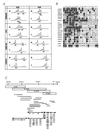
Figure 1
Figure 1
LOH at human chromosome 8p in primary esophageal cancer samples and genomic contigs at 8p22. (A) Representative LOH analysis from two cases, E26 and E46. Fluorescent PCR products from normal (N) and tumor (T) DNAs were analyzed. The x axis shows DNA fragment size in bp (depicted in boxes, compared with size markers). Patient E26 was informative for all four markers; LOH for D8S264, LPL, and D8S136 and allelic retention in FGFR1 (because a 167-bp peak in tumor is more than 50% of that in normal). Patient E46 was informative at D8S264 and D8S136, and one allele at each was lost, whereas LPL and FGFR1 loci were homozygous. (B) Summary of LOH analyses. All the patients with loss at least at one locus are shown. The closed circles indicate loss of an allele, the circles with crosses indicate noninformation because of homozygosity, and the open circles depict the retention of both alleles. The dark hatched areas indicate regions of allele loss, whereas the light hatched areas show regions of noninformation within allele loss areas. Further studies focused on the region near the marker D8S261 locus shown in a boxed area. The column numbers correspond to patients, whereas the row numbers indicate polymorphic markers. (C) Genomic contigs at 8p22. The upper line depicts the location of polymorphic loci on 8p. YAC (boxes) and BAC (horizontal lines) contigs were constructed. The cDNA selection was performed on three YAC DNAs (triangles). The shotgun sequencing were carried out on BACs (triangles). Eighty-seven potentially expressed sequences were isolated and located within the contigs, which are shown in the lower portion. The underlined characters are the sequences that are expressed in normal tissues. A candidate fragment, e37, corresponds to the F37 cDNA described in text. Areas with arrows show locations of candidate cDNAs. After expression analysis in tumor and normal tissues, nine cDNAs (circled characters) were subjected to partial cloning and further analysis.
Figure 2
Figure 2
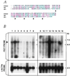
The predicted FEZ1 amino acid sequence and the expression. (A) Comparison of the predicted amino acid sequences corresponding to the DNA-binding and leucine-zipper regions. Comparison of Fez1 (amino acids 301–369 are depicted) with Atf-5 and KIAA0522 proteins are shown. Identical (red shading) or similar (blue shading) residues among Fez1 and the other two proteins are indicated. Gaps introduced by the fasta program are represented by spaces. Closed circles denote repeated leucine residues. (B) FEZ1 gene expression. Northern blot analysis of normal tissue RNAs. Probes used were the FEZ1 ORF probe (upper) and the control β-actin probe (lower). Poly(A)+ RNA (5 μg) was loaded. The arrowhead on the left indicates the 6.8-kb FEZ1 transcript. RNAs in lanes were from: 1, heart; 2, brain; 3, placenta; 4, lung; 5, liver; 6, skeletal muscle; 7, kidney; 8, pancreas; 9, spleen, 10, thymus; 11, prostate; 12, testes; 13, ovary; 14, small intestine; 15, colon; 16, peripheral blood lymphocyte.
Figure 3
Figure 3
Alterations of FEZ1 in tumors. (A) FEZ1 gene expression in cancers. Probes used were a FEZ1 cDNA probe (Upper) and a control β-actin probe (Lower). Poly(A)+ RNA (5 μg) from cancer cell lines was loaded. The arrowhead on the left indicates the 6.8-kb transcript of FEZ1. RNAs were from: esophageal cancer cell lines KYSE170 (lane 1), TE12 (lane 2), TE8 (lane 3), and TE3 (lane 4); prostate cancer cell lines DU145 (lane 5), LNCaP (lane 6), PC3 (lane 7), and normal prostate (lane 8); breast cancer cell lines MB231 (lane 9), SKBr3 (lane 10), BT549 (lane 11), HBL100 (lane 12), MB436 (lane 13), BT20 (lane 14), MB543 (lane 15), MB175 (lane 16), MCF7 (lane 17), and T47B (lane 18); normal breast (lane 19); and total RNA of normal breast (lane 20); promyelocytic leukemia cell line HL60 (lane 21); cervical cancer cell line HeLa S3 (lane 22); chronic myelogenous leukemia cell line K562 (lane 23); lymphoblastic leukemia cell line MOLT4 (lane 24); Burkitt’s lymphoma cell line Raji (lane 25); colorectal adenocarcinoma cell line SW480 (lane 26); lung cancer cell line A549 (lane 27); and melanoma cell line G361 (lane 28). (B) Sequence chromatograms of three mutations. A point mutation (TCC/Ser → CCC/Pro) at codon 29 was found (T) in an primary esophageal cancer, E44. As controls, normal sequences from normal DNA from patient E44 and BAC sequence (N) were analyzed, both producing the same result. A bold line indicates the altered codon. In primary esophageal cancer, E50, a point mutation (AAG/Lys → GAG/Glu) at codon 119 was found (T). The normal BAC sequence is shown in (N). The point mutation (CAG/Gln → TAG/STOP) at codon 501 was observed in prostate cancer PC3 cells (T). Note that the sequence chromatogram from this cell line is shown in the 3′ to 5′ direction. Repeated sequencing showed a weak signal corresponding to guanine (G) within a large adenine (A) signal in the first nucleotide at codon 501, suggesting the retention of the normal allele in a fraction of the cancer cells. Expression analysis with RT-PCR detected only the mutated transcript in this cell line. The normal BAC sequence is shown in (N). (C) Truncated FEZ1 transcripts observed in cancers. The normal exon/intron structure (top drawing) was determined by sequencing of brain, prostate, and esophagus cDNAs and by sequencing FEZ1 gene in BAC. The boxes indicate exons, and the shaded areas show the ORF of 1,788 bp. The introns are depicted by horizontal lines. Aberrant transcripts observed in tumors are depicted in bold lines. The location of mutations are shown as closed circles. LZ, leucine-zipper motif; FS, frameshift. (D) Southern blot analysis of the FEZ1 gene locus. High-Mr DNAs from cancer cells were cleaved with EcoRI, separated electrophoretically, transferred to a nylon membrane, and probed with the 1.7-kb FEZ1 ORF probe. DNAs (10 μg/lane) were from lanes: 1, MB436S; 2, normal; 3, MB231; 4, MB361; 5, TE8; 6, TE3; 7, normal (different origin from normal DNA in lane 2).

Similar articles

Cited by

References

    1. Cavenee W, Dryja T P, Phillips R A, Benedict W F, Godbout R, Gallie B L, Murphree A L, Strong L C, White R L. Nature (London) 1983;305:779–784. - PubMed
    1. Lasko D, Cavenee W. Annu Rev Genet. 1991;25:281–314. - PubMed
    1. Weinberg R. Science. 1991;254:1138–1146. - PubMed
    1. Knudson A. Proc Natl Acad Sci USA. 1993;90:10914–10921. - PMC - PubMed
    1. Nowell P. Adv Cancer Res. 1993;62:1–17. - PubMed

Publication types

MeSH terms