. 1999 Mar 30;96(7):3934-9.
doi: 10.1073/pnas.96.7.3934.

Specific association of the gene product of PKD2 with the TRPC1 channel

Affiliations
Free PMC article

Specific association of the gene product of PKD2 with the TRPC1 channel

L Tsiokas et al. Proc Natl Acad Sci U S A. .
Free PMC article

Abstract

The function(s) of the genes (PKD1 and PKD2) responsible for the majority of cases of autosomal dominant polycystic kidney disease is unknown. While PKD1 encodes a large integral membrane protein containing several structural motifs found in known proteins involved in cell-cell or cell-matrix interactions, PKD2 has homology to PKD1 and the major subunit of the voltage-activated Ca2+ channels. We now describe sequence homology between PKD2 and various members of the mammalian transient receptor potential channel (TRPC) proteins, thought to be activated by G protein-coupled receptor activation and/or depletion of internal Ca2+ stores. We show that PKD2 can directly associate with TRPC1 but not TRPC3 in transfected cells and in vitro. This association is mediated by two distinct domains in PKD2. One domain involves a minimal region of 73 amino acids in the C-terminal cytoplasmic tail of PKD2 shown previously to constitute an interacting domain with PKD1. However, distinct residues within this region mediate specific interactions with TRPC1 or PKD1. The C-terminal domain is sufficient but not necessary for the PKD2-TRPC1 association. A more N-terminal domain located within transmembrane segments S2 and S5, including a putative pore helical region between S5 and S6, is also responsible for the association. Given the ability of the TRPC to form functional homo- and heteromultimeric complexes, these data provide evidence that PKD2 may be functionally related to TRPC proteins and suggest a possible role of PKD2 in modulating Ca2+ entry in response to G protein-coupled receptor activation and/or store depletion.


Figures

Figure 1
Figure 1
Sequence alignment of the TRP-like regions in human PKD2 (hPKD2, GenBank accession no. U50928), hPKD2b (AF092170), hTRPC1 (Z73903), bovine TRPC4 (bTRPC4, X99792) mouse TRPC5 (mTRPC5, AJ006204), hTRPC3 (Y13758), and mTRPC6 (U49069). The first and last amino acids of each sequence, numbered according to their corresponding cDNAs, are shown in the end of each sequence. Identical or similar substitutions (similar substitutions are grouped as follows: F and Y; I and V; R and K; L and M; or N, D, Q, and E) conserved in five or seven sequences are shown in gray or by a dot on top of the sequences, respectively. Low consensus shown in gray allows the identification of similar residues within the TRP family. Alignment was done according to hierarchical clustering using a blosum62 scoring table (35). Amino acid residues corresponding to putative TM segments (S3–S6) are underlined, and the boxed area shows a putative pore helical region (30). Asterisks denote conserved residues that may participate in the formation of the ion pore.
Figure 2
Figure 2
Coimmunoprecipitation of HA-PKD2 and M-TRPC1. HEK293T cells were cotransfected with the indicated combinations of expression plasmids. HA-PKD2 was immunoprecipitated (IP) with α-HA and immunocomplexes were analyzed by immunoblotting (IB) with α-myc (Upper). Expression levels of myc-tagged fusions (M-TRPC1, TRPC3-M, and α1c-M) in the precleared lysates of HEK293T cells before immunoprecipitation are shown (Lower). The positions of molecular mass markers in kDa are shown (Left).
Figure 3
Figure 3
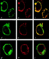
Colocalization of HA-PKD2 and TRPC1 in live cells. HEK293T cells were cotransfected with HA-PKD2 and F-TRPC1 (a–c), HA-PKD2 and M-TRPC1 (d–f), or HA-PKD2 and TRPC3-M (gi), and the subcellular distribution of the encoded proteins was determined by double immunofluorescence using confocal microscopy. FLAG- or myc-tagged proteins were stained with a FITC-conjugated secondary antibody, while HA-PKD2 was stained with rhodamine-conjugated secondary antibody. Computerized images of green fluorescein staining corresponding to F-TRPC1, M-TRPC1, or TRPC3-M are shown in a, d, and g, respectively, whereas red rhodamine images corresponding to HA-PKD2 are shown in b, e, and h. Fluorescein and rhodamine merged images corresponding to HA-PKD2 and F-TRPC1, HA-PKD2 and M-TRPC1, or HA-PKD2 and TRPC3-M subcellular distributions are shown in c, f, and i, respectively.
Figure 4
Figure 4
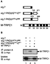
Coimmunoprecipitation of membrane bound versions of the C-terminal cytoplasmic region of PKD2 and M-TRPC1. (A) Diagrammatic representation of constructs used in (B). The sIg.7 cassette contains the leader sequence of CD5 (░⃞) fused to the CH2-CH3 domain of human IgG1 followed by the TM region of CD7 (■). sIg.7-PKD2/Q743–E871 or sIg.7-PKD2/I679–V968 were made by fusing a subregion (743–871) or the entire C-terminal cytoplasmic tail of PKD2 (679–968) to the C terminus of sIg.7, respectively. Numbering was done according to full-length PKD2 (U50928). Schematic representation of M-TRPC1 including all six TM segments (S1–S6) is also shown. (B) HEK293T cells were cotransfected with sIg.7 (lane 1), sIg.7-PKD2/I679-V968 (lane 2), or sIg.7-PKD2/Q743-E871 (lane 3) and M-TRPC1 (lanes 1–3). sIg.7 fusions were immunoprecipitated (IP) with protein A and immunocomplexes (Upper) or lysates from transiently transfected cells (Lower) were determined by immunoblotting (IB) and probed with α-myc.
Figure 5
Figure 5
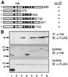
PKD2-TRPC1 association via a TM region in PKD2. (A) Schematic representation of wild type (1–968; HA-PKD2) or truncation mutants (HA-PKD2/1–379, 1–643, 1–742, 1–871, and 1–702) of HA-PKD2 and summary of results shown in B. Black boxes (■) represent TM segments S1–S6. (B) HEK293T cells were cotransfected with FLAG-tagged bacterial alkaline phosphatase and HA-PKD2 (lane 1), pCDNA3 vector and F-TRPC1 (lane 2), F-TRPC1 and HA-PKD2 (lane 3), HA-PKD2/1–379 (lane 4), HA-PKD2/1–643 (lane 5), HA-PKD2/1–742 (lane 6), HA-PKD2/1–871 (lane 7), or HA-PKD2/1–702 (lane 8). Cells were lysed in IP-500 and HA-tagged proteins were immunoprecipitated (IP) with α-HA. Immunocomplexes were determined by immunoblotting (IB) with α-FLAG (Upper). Expression levels of HA- or FLAG-tagged proteins are shown (Middle or Lower, respectively).
Figure 6
Figure 6
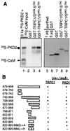
Identification of the C-terminal interacting domain in PKD2. (A) Direct interaction between the C-terminal tail of PKD2 and TRPC1. In vitro column binding of 35S-PKD2 and 35S-CaM to GST-TRPC1/D639–S750 or GST-TRPC1/S662–S750 (lanes 1–4). Lane 1 shows 0.1× the input amount of 35S-PKD2 and 35S-CaM. Bound 35S-PKD2 and 35S-CaM to GST, GST-TRPC1/D639-S750, or GST-TRPC1/S662–S750 is shown in lanes 2–4 and the immobilized amounts of GST, GST-TRPC1/D639–S750, and GST-TRPC1/S662–S750 subjected to in vitro column binding are shown in lanes 7–9. To obtain an estimate of the immobilized amounts of GST, GST-TRPC1/D639–S750, or GST-TRPC1/S662–S750 used in the in vitro column binding assays, 1 or 5 μg of purified GST is shown in lanes 5 and 6. A lower molecular weight band corresponding to a C-terminal proteolytic product of 35S-PKD2 is shown by an arrow below the band corresponding to full-length 35S-PKD2. (B) cDNAs corresponding to the entire C-terminal cytoplasmic region of PKD2, systematic N- or C-terminal deletions of this region (amino acid residues 679–968) or nonconserved substitutions in the region 822–895 were tested for their interaction with the cytoplasmic tail of TRPC1 (D639–S750) or PKD1 (P4124–T4303) by the yeast two-hybrid assay. Positive interactions were scored for both survival in plates lacking histidine (his+) and production of β-galactosidase (lacZ+).

Similar articles

Cited by

References

    1. Gabow P A. N Engl J Med. 1993;329:332–342. - PubMed
    1. The European Polycystic Kidney Disease Consortium. Cell. 1994;77:881–894. - PubMed
    1. The International Polycystic Kidney Disease Consortium. Cell. 1995;81:289–298. - PubMed
    1. Mochizuki T, Wu G, Hayashi T, Xenophontos S L, Veldhuisen B, Saris J J, Reynolds D M, Cai Y, Gabow P A, Pierides A, et al. Science. 1996;272:1339–1342. - PubMed
    1. Sandford R, Sgotto B, Aparicio S, Brenner S, Vaudin M, Wilson R K, Chissoe S, Pepin K, Bateman A, Chothia C, Hughes J, Harris P. Hum Mol Genet. 1997;6:1483–1489. - PubMed

Publication types

MeSH terms

Associated data