

Induction of solid tumor differentiation by the peroxisome proliferator-activated receptor-gamma ligand troglitazone in patients with liposarcoma
- PMID: 10097144
- PMCID: PMC22401
- DOI: 10.1073/pnas.96.7.3951
Induction of solid tumor differentiation by the peroxisome proliferator-activated receptor-gamma ligand troglitazone in patients with liposarcoma
Abstract
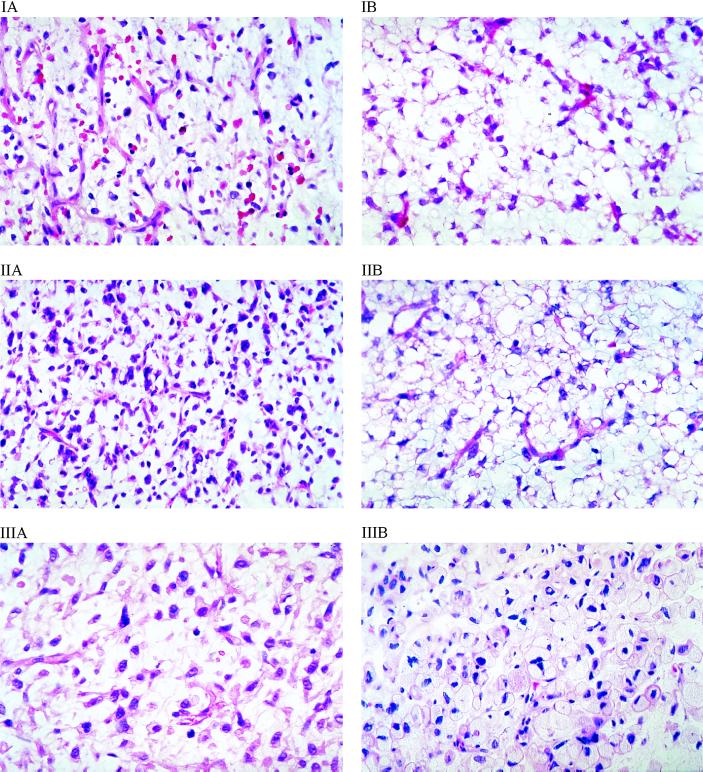
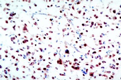
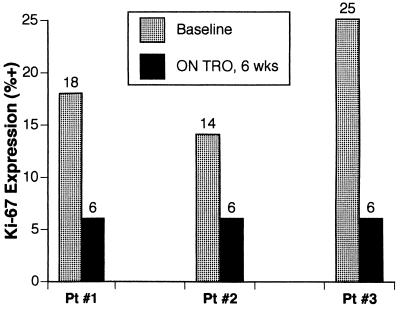
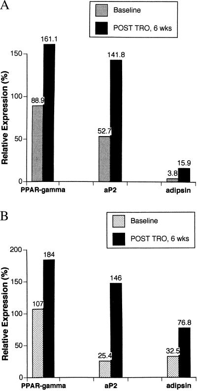
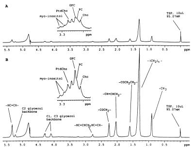
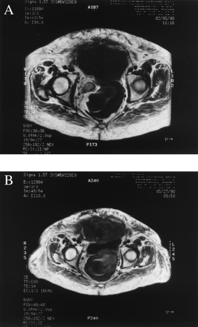
Agonist ligands for the nuclear receptor peroxisome proliferator-activated receptor-gamma have been shown to induce terminal differentiation of normal preadipocytes and human liposarcoma cells in vitro. Because the differentiation status of liposarcoma is predictive of clinical outcomes, modulation of the differentiation status of a tumor may favorably impact clinical behavior. We have conducted a clinical trial for treatment of patients with advanced liposarcoma by using the peroxisome proliferator-activated receptor-gamma ligand troglitazone, in which extensive correlative laboratory studies of tumor differentiation were performed. We report here the results of three patients with intermediate to high-grade liposarcomas in whom troglitazone administration induced histologic and biochemical differentiation in vivo. Biopsies of tumors from each of these patients while on troglitazone demonstrated histologic evidence of extensive lipid accumulation by tumor cells and substantial increases in NMR-detectable tumor triglycerides compared with pretreatment biopsies. In addition, expression of several mRNA transcripts characteristic of differentiation in the adipocyte lineage was induced. There was also a marked reduction in immunohistochemical expression of Ki-67, a marker of cell proliferation. Together, these data indicate that terminal adipocytic differentiation was induced in these malignant tumors by troglitazone. These results indicate that lineage-appropriate differentiation can be induced pharmacologically in a human solid tumor.
Figures
Similar articles
-
Biochemical correlates of thiazolidinedione-induced adipocyte differentiation by high-resolution magic angle spinning NMR spectroscopy.Magn Reson Med. 2002 Oct;48(4):602-10. doi: 10.1002/mrm.10256. Magn Reson Med. 2002. PMID: 12353276
-
Troglitazone improves psoriasis and normalizes models of proliferative skin disease: ligands for peroxisome proliferator-activated receptor-gamma inhibit keratinocyte proliferation.Arch Dermatol. 2000 May;136(5):609-16. doi: 10.1001/archderm.136.5.609. Arch Dermatol. 2000. PMID: 10815854 Clinical Trial.
-
Terminal differentiation of human liposarcoma cells induced by ligands for peroxisome proliferator-activated receptor gamma and the retinoid X receptor.Proc Natl Acad Sci U S A. 1997 Jan 7;94(1):237-41. doi: 10.1073/pnas.94.1.237. Proc Natl Acad Sci U S A. 1997. PMID: 8990192 Free PMC article.
-
Peroxisome proliferator-activated receptor gamma (PPargamma) as a novel target for prostate cancer.Invest New Drugs. 2002 May;20(2):195-200. doi: 10.1023/a:1015670126203. Invest New Drugs. 2002. PMID: 12099579 Review.
-
Troglitazone and related compounds: therapeutic potential beyond diabetes.Life Sci. 2000 Oct 6;67(20):2405-16. doi: 10.1016/s0024-3205(00)00829-8. Life Sci. 2000. PMID: 11065164 Review.
Cited by
-
Combined COX-2/PPARγ Expression as Independent Negative Prognosticator for Vulvar Cancer Patients.Diagnostics (Basel). 2021 Mar 10;11(3):491. doi: 10.3390/diagnostics11030491. Diagnostics (Basel). 2021. PMID: 33802010 Free PMC article.
-
Association between longer therapy with thiazolidinediones and risk of bladder cancer: a cohort study.J Natl Cancer Inst. 2012 Sep 19;104(18):1411-21. doi: 10.1093/jnci/djs328. Epub 2012 Aug 9. J Natl Cancer Inst. 2012. PMID: 22878886 Free PMC article.
-
Chemotherapy and chemoprevention by thiazolidinediones.Biomed Res Int. 2015;2015:845340. doi: 10.1155/2015/845340. Epub 2015 Mar 19. Biomed Res Int. 2015. PMID: 25866814 Free PMC article. Review.
-
Peroxisome proliferator-activated receptor gamma regulates E-cadherin expression and inhibits growth and invasion of prostate cancer.Mol Cell Biol. 2006 Oct;26(20):7561-74. doi: 10.1128/MCB.00605-06. Mol Cell Biol. 2006. PMID: 17015477 Free PMC article.
-
Clinical Efficacy of a Novel Therapeutic Principle, Anakoinosis.Front Pharmacol. 2018 Nov 28;9:1357. doi: 10.3389/fphar.2018.01357. eCollection 2018. Front Pharmacol. 2018. PMID: 30546308 Free PMC article. Review.
References
-
- Warrell R P, Frankel S R, Miller W H, Scheinberg D A, Itri L M, Hittleman W N, Vyas R, Andreef M, Tafuri A, Jakubowski A, et al. N Engl J Med. 1991;324:1385–1393. - PubMed
-
- Warrell R P, de The H, Wang Z-Y, Degos L. N Engl J Med. 1993;329:177–189. - PubMed
-
- Tontonoz P, Hu E, Graves R A, Budavari A I, Spiegelman B M. Genes Dev. 1994;8:1224–1234. - PubMed
-
- Tontonoz P, Hu E, Spiegelman B M. Cell. 1994;79:1147–1156. - PubMed
Publication types
MeSH terms
Substances
Grants and funding
LinkOut - more resources
Full Text Sources
Other Literature Sources
Medical
