. 1999 Mar 30;96(7):3957-62.
doi: 10.1073/pnas.96.7.3957.

Evidence for a structural motif in toxins and interleukin-2 that may be responsible for binding to endothelial cells and initiating vascular leak syndrome

Affiliations
Free PMC article

Evidence for a structural motif in toxins and interleukin-2 that may be responsible for binding to endothelial cells and initiating vascular leak syndrome

R Baluna et al. Proc Natl Acad Sci U S A. .
Free PMC article

Abstract

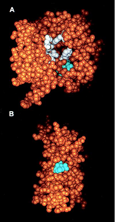
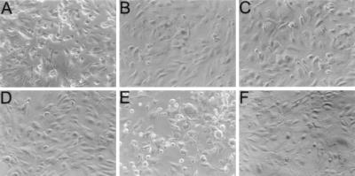
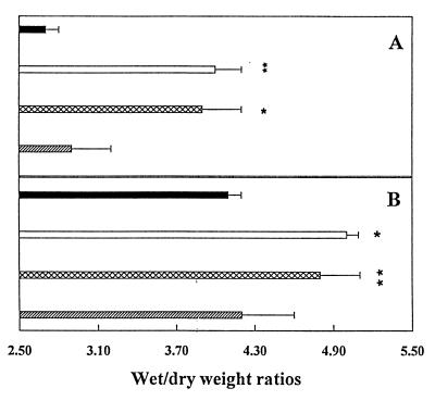
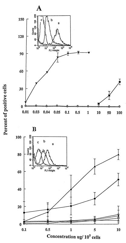
The dose-limiting toxicity of interleukin-2 (IL-2) and immunotoxin (IT) therapy in humans is vascular leak syndrome (VLS). VLS has a complex etiology involving damage to vascular endothelial cells (ECs), extravasation of fluids and proteins, interstitial edema, and organ failure. IL-2 and ITs prepared with the catalytic A chain of the plant toxin, ricin (RTA), and other toxins, damage human ECs in vitro and in vivo. Damage to ECs may initiate VLS; if this damage could be avoided without losing the efficacy of ITs or IL-2, larger doses could be administered. In this paper, we provide evidence that a three amino acid sequence motif, (x)D(y), in toxins and IL-2 damages ECs. Thus, when peptides from RTA or IL-2 containing this sequence motif are coupled to mouse IgG, they bind to and damage ECs both in vitro and, in the case of RTA, in vivo. In contrast, the same peptides with a deleted or mutated sequence do not. Furthermore, the peptide from RTA attached to mouse IgG can block the binding of intact RTA to ECs in vitro and vice versa. In addition, RTA, a fragment of Pseudomonas exotoxin A (PE38-lys), and fibronectin also block the binding of the mouse IgG-RTA peptide to ECs, suggesting that an (x)D(y) motif is exposed on all three molecules. Our results suggest that deletions or mutations in this sequence or the use of nondamaging blocking peptides may increase the therapeutic index of both IL-2, as well as ITs prepared with a variety of plant or bacterial toxins.


Figures

Figure 1
Figure 1
Localization of the LDV and LDL sequences in RTA (A) and IL-2 (B). Space filling models of the three-dimensional structures of RTA (A) (PDB accession no. 1br5.pdb) and IL-2 (B) (PDB accession no. 1irl.pdb) are shown with the atoms of the LDV residues of RTA and the LDL residues of IL-2 shown in cyan, the active site residues of RTA (Y80, Y123, E177, R180, N209, and W211) in white, and all other atoms in orange. Models were generated with the insight ii program (Micron Separations).
Figure 2
Figure 2
Effect of RFB4 peptides on the morphology of HUVEC monolayers. HUVEC monolayers were incubated at 37°C for 18 hr with 10−6 M RFB4-LDV+ (A), RFB4-LDV (B), RFB4-GQT (C), RFB4-LDL+ (E), RFB4-LDL (F), or medium only (D) and then examined by phase-contrast microscopy (magnification ×20). Normal monolayers consisted of highly packed cells with elongated shapes (BD and F), whereas damaged cells were rounded up and detached from the plate (A and E).
Figure 3
Figure 3
The in vivo effect of RFB4-RTA peptides. (A) SCID mice with human xenografts were injected with 200 μg of RFB4-dgRTA (□), RFB4-LDV+ (⊠), RFB4-GQT (▨), or saline (■), and the wet/dry weight ratios of the human skin were determined. (B) SCID mice were injected as described in A and the wet/dry weight ratios of lungs were determined. Values represent the mean of three experiments ± SD. Asterisks indicate a significant difference from saline-treated mice (∗, P < 0.02; ∗∗, P < 0.01).
Figure 4
Figure 4
Binding of dgRTA, PE38-lys, and RFB4 peptides to HUVECs. HUVECs (105) were incubated on ice for 30 min with FITC reagents in 100 μl PBS/BSA/AZ at varying concentrations, washed, fixed in 1% paraformaldehyde, and analyzed by flow cytometry. Values represent the mean ± SD of three experiments. (A) FITC-dgRTA (▾), FITC-PE38-lys (■), FITC-carbonic anhydrase (control) (○). Inset shows the histograms of flow cytometric analyses of the binding of (a) dgRTA, (b) PE38-lys, and (c) carbonic anhydrase to HUVECs. (B) FITC-RFB4-LDV+ (▴), FITC-RFB4-LDV (▵), FITC-RFB4-GQT+ (◊), FITC-RFB4 (□), FITC-RFB4-LDL+ (●), FITC-RFB4-LDL (⧫). Inset shows the histograms of (a) RFB4-LDV+, (b) RFB4-LDL+, and (c) RFB4.
Figure 5
Figure 5
Inhibition of the binding of dgRTA and RFB4-LDV+ to HUVECs. HUVECs (105) were incubated on ice for 30 min with FITC-dgRTA (A) or FITC-RFB4-LDV+ (B) in the presence or absence of 100-fold excess of dgRTA (■), RFB4-LDV+ (⊠), RFB4 (░⃞), Fn (▨), or PE38-lys (□) in 100 μl PBS/BSA/AZ. The percent inhibition of binding to HUVECs is presented. Values represent the means ± SD of three experiments.

Similar articles

Cited by

References

    1. Vitetta E S, Thorpe P E, Uhr J W. Immunol Today. 1993;14:252–259. - PubMed
    1. Sausville E A, Vitetta E S. In: Monoclonal Antibody-Based Therapy of Cancer. Grossbard M L, editor. Vol. 4. Basel: Marcel Dekker; 1997. pp. 81–89.
    1. Baluna R, Vitetta E S. Immunopharmacology. 1996;37:117–132. - PubMed
    1. Engert A, Sausville E A, Vitetta E S. In: Clinical Applications of Immunotoxins. Frankel A E, editor. Vol. 2. Berlin: Springer; 1997. pp. 13–33.
    1. Dutcher J P, Gaynor E R, Boldt D H, Doroshow J H, Bar M H, Sznol M, Mier J, Sparano J, Fisher R I, Weiss G, et al. J Clin Oncol. 1991;9:641–648. - PubMed

MeSH terms

LinkOut - more resources