. 1999 Mar 30;96(7):3987-92.
doi: 10.1073/pnas.96.7.3987.

Ectopic expression of prion protein (PrP) in T lymphocytes or hepatocytes of PrP knockout mice is insufficient to sustain prion replication

Affiliations

Ectopic expression of prion protein (PrP) in T lymphocytes or hepatocytes of PrP knockout mice is insufficient to sustain prion replication

A J Raeber et al. Proc Natl Acad Sci U S A. .

Abstract

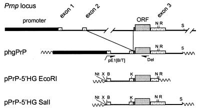
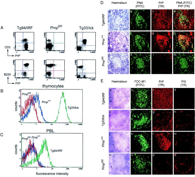
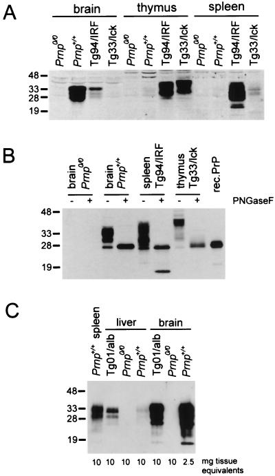
The cellular form of the Prion protein (PrPC) is necessary for prion replication in mice. To determine whether it is also sufficient, we expressed PrP under the control of various cell- or tissue-specific regulatory elements in PrP knockout mice. The interferon regulatory factor-1 promoter/Emu enhancer led to high PrP levels in the spleen and low PrP levels in the brain. Following i.p. scrapie inoculation, high prion titers were found in the spleen but not in the brain at 2 weeks and 6 months, showing that the lymphoreticular system by itself is competent to replicate prions. PrP expression directed by the Lck promoter resulted in high PrP levels on T lymphocytes only but, surprisingly, did not allow prion replication in the thymus, spleen, or brain following i.p. inoculation. A third transgenic line, which expressed PrP in the liver under the control of the albumin promoter/enhancer-albeit at low levels-also failed to replicate prions. These results show that expression of PrP alone is not sufficient to sustain prion replication and suggest that additional components are needed.


Figures

Figure 1
Figure 1
Schematic representation of half-genomic PrP transgenes driven by heterologous promoters. The genomic mouse Prnp locus is shown on top (37). Construction of the “half-genomic” PrP vector (phgPrP) lacking the 12-kb intron 2 has been described (14). Using PCR with the primers PE1 and Del, a BamHI site was introduced at the 5′ end of exon 1 in phgPrP. The resulting promoterless construct pPrP-5′HG EcoRI was cloned into Bluescript, and the PrP sequence was extended up to the SalI site in the 3′ noncoding region by introducing the NarI-SalI fragment of phgPrP to yield pPrP-5′HG SalI. Promoter cassettes were inserted into the BamHI site of pPrP-5′HG SalI to yield plck-PrP-5′HG SalI, pEμ/IRF1-PrP-5′HG SalI, and pAlbumin-PrP-5′HG SalI. B, BamHI; K, KpnI; N, NarI; Nt, NotI; R, EcoRI; S, SalI; X, XbaI. Wavy lines, vector sequences.
Figure 2
Figure 2
Analysis of PrP expression by FACS and immunohistochemistry. FACS analysis for cell-surface PrP was carried out on (A) splenocytes, (B) thymocytes, and (C) peripheral blood leukocytes (PBL) gated for lymphocytes from Prnp+/+, Prnp0/0, Tg94/IRF, and Tg33/lck mice. Cells were stained with anti-PrP polyclonal antisera R340 and phycoerythrin-conjugated anti-rabbit IgG, and analyzed by FACS gated for lymphocytes. (A) For two-color FACS analysis, PrP staining was followed by B cell staining with FITC-conjugated anti-B220 antibodies or T cell staining with FITC-conjugated anti-CD3 antibodies. (D) Double immunofluorescence analysis of splenic germinal centers in noninoculated Tg94/IRF (a–d), wild-type mice (e–h), and Prnp0/0 mice (j–m). Sections were stained with haemalaun (a, e, j), with peanut agglutinin (PNA; green; b, f, k), and with antiserum R340 to PrP (Texas red; c, g, l). The majority of B cells PNA labeled in the germinal center were PrP-positive in Tg94/IRF mice (yellow signal in superimposed images; d) and in wild-type mice (h), but PrP-negative in Prnp0/0 mice (m). (Original magnification ×250.) (E) Immunofluorescence labeling of FDCs and PrP on consecutive sections of spleen from noninoculated Tg94/IRF (a–d), Tg33/lck (e–h), wild-type (j–m), and Prnp0/0 mice (n–q). Sections were stained with haemalaun (a, e, j, n), antibody FDC-M1 to FDC (green; b, f, k, o), antiserum R340 to PrP (Texas red; c, g, l, p), and rabbit pre-immune serum (PIS; d, h, m, q). In wild-type spleens (k, l), PrP was stained exclusively in the germinal centers, most strongly in the areas also stained by FDC-M1. In Tg94/IRF mice (b, c), PrP was evenly distributed over the entire section, including the region also stained by FDC-M1. In Tg33/lck spleens, PrP was visualized mainly in the T cell areas, but some cells were stained in the region also stained by FDC-M1. No PrP staining above background (q) was found in germinal centers of Prnp0/0 mice (p). (Original magnification ×250.)
Figure 3
Figure 3
Immunoblot analysis for PrP in tissues of various mouse lines. (A) Aliquots (120 μg of protein) of tissue homogenates as indicated were loaded per lane. (B) Aliquots (40 μg of protein) of tissue homogenates were digested with 500 units of N-glycosidase F for 2 h at 37°C. (C) Aliquots of tissue homogenates as indicated were immunoprecipitated with 6H4 monoclonal antibody coupled to Sepharose 4B. The eluted proteins were subjected to Western blotting and PrP was detected on blots with 1:10,000 diluted polyclonal anti-PrP antiserum 1B3. Molecular mass markers are indicated on the left in kDa.

References

    1. Büeler H, Aguzzi A, Sailer A, Greiner R A, Autenried P, Aguet M, Weissmann C. Cell. 1993;73:1339–1347. - PubMed
    1. Eklund C M, Kennedy R C, Hadlow W J. J Infect Dis. 1967;117:15–22. - PubMed
    1. Fraser H, Dickinson A G. Nature (London) 1970;226:462–463. - PubMed
    1. Kitamoto T, Muramoto T, Mohri S, Dohura K, Tateishi J. J Virol. 1991;65:6292–6295. - PMC - PubMed
    1. Muramoto T, Kitamoto T, Tateishi J, Goto I. Am J Pathol. 1992;140:1411–1420. - PMC - PubMed

Publication types