. 1999 Mar 30;96(7):4034-9.
doi: 10.1073/pnas.96.7.4034.

Segregating the functions of human hippocampus

Affiliations

Segregating the functions of human hippocampus

B A Strange et al. Proc Natl Acad Sci U S A. .

Abstract

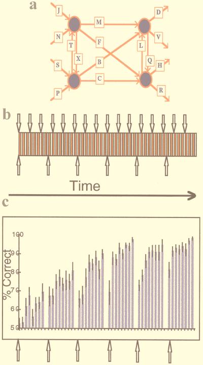
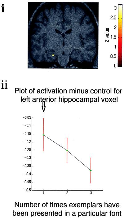
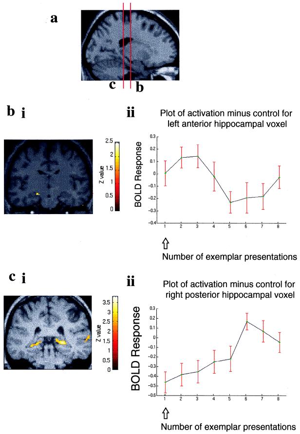
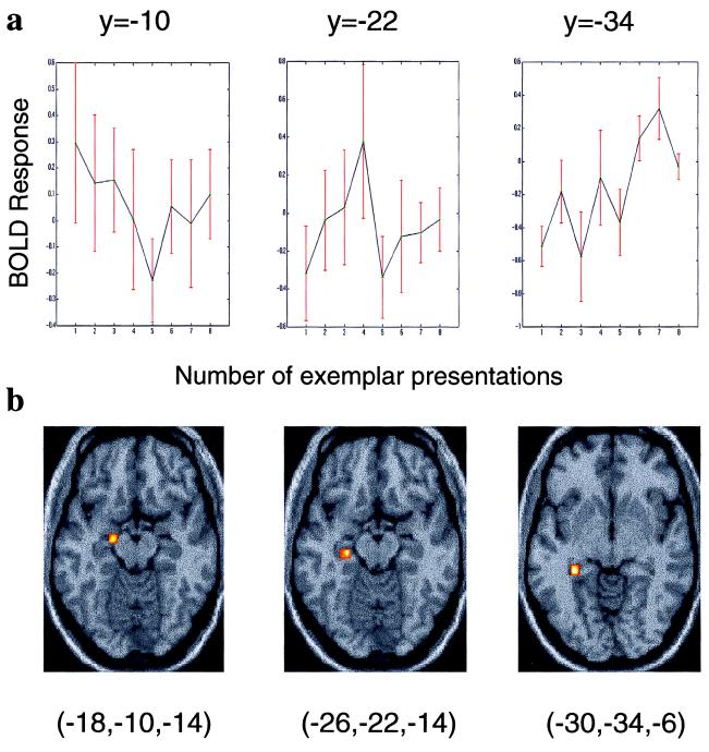
It is now accepted that hippocampal lesions impair episodic memory. However, the precise functional role of the hippocampus in episodic memory remains elusive. Recent functional imaging data implicate the hippocampus in processing novelty, a finding supported by human in vivo recordings and event-related potential studies. Here we measure hippocampal responses to novelty, using functional MRI (fMRI), during an item-learning paradigm generated from an artificial grammar system. During learning, two distinct types of novelty were periodically introduced: perceptual novelty, pertaining to the physical characteristics of stimuli (in this case visual characteristics), and exemplar novelty, reflecting semantic characteristics of stimuli (in this case grammatical status within a rule system). We demonstrate a left anterior hippocampal response to both types of novelty and adaptation of these responses with stimulus familiarity. By contrast to these novelty effects, we also show bilateral posterior hippocampal responses with increasing exemplar familiarity. These results suggest a functional dissociation within the hippocampus with respect to the relative familiarity of study items. Neural responses in anterior hippocampus index generic novelty, whereas posterior hippocampal responses index familiarity to stimuli that have behavioral relevance (i.e., only exemplar familiarity). These findings add to recent evidence for functional segregation within the human hippocampus during learning.


Figures

Figure 1
Figure 1
The introduction of perceptual and exemplar novelty in the context of item learning. (a) The artificial grammar system. Grammatical four-consonant letter strings are formed by starting on the left and moving right in the direction of the arrows. (b) Experimental design. Every exemplar-set presentation constitutes an activation epoch (red rectangle), each of which is followed by a control epoch (white rectangle). Every exemplar set is presented eight times, after which a new set of exemplars is presented, allowing the introduction of exemplar novelty (↑). Every three activation epochs the font in which exemplar strings are presented is changed, enabling the introduction of perceptual novelty (↓). (c) Behavioral data averaged for all subjects expressed as a percentage of correct grammaticality judgements (error bars here, and in all subsequent plots, depict ±1 SE). The data show improving grammaticality judgements as subjects become increasingly familiar with each set of exemplars. When a new exemplar set is presented, performance falls, but across the entire experiment subjects gradually acquire more abstract knowledge about the grammar system and use this knowledge to maintain performance after a change in exemplar set.
Figure 2
Figure 2
Hippocampal region in which there is a significant time × condition interaction in response to perceptual novelty and adaptation with familiarity. (i) Coronal section of a T1-weighted anatomical image (at y = −16) that conforms to the stereotaxic space. The image is taken from 1 of the 14 subjects. T1-weighted images in all subsequent figures are taken from the same subject. Superimposed on this section is a SPM (thresholded at P < 0.01) indicating a decreasing linear time × condition interaction in the left hippocampus after introduction of novel font. The section has been chosen to demonstrate left anterior hippocampal activation (x, y, z coordinates −22, −16, −24; Z = 3.25). (ii) Graphic representation of activation at this voxel relative to the baseline condition as a function of repeated presentation of fonts. The plotted time course shows the BOLD response collapsed across the 16 font changes and averaged across all subjects. ↓, introduction of novel font.
Figure 3
Figure 3
Dissociation in the anterior–posterior hippocampal axis for exemplar novelty. (a) T1-weighted anatomical sagittal image with red lines indicating the anterior–posterior positions of the coronal sections demonstrated in b and c. (bi) SPM prepared as above showing enhanced left anterior hippocampal activation after the introduction of exemplar novelty followed by deactivation (threshold P < 0.05). The coronal section has been chosen to demonstrate left anterior hippocampal activation (coordinates −18, −16, −14; Z = 2.60). (bii) Graphic representation of activation at this voxel relative to the baseline condition against number of presentations of an exemplar set. The plot shows the BOLD response collapsed across the six exemplar changes and averaged across all subjects. ↑, introduction of novel exemplar set. (ci) SPM (threshold P < 0.01) showing that increasing familiarity with exemplars activates the posterior hippocampus bilaterally. The coronal section has been chosen to demonstrate right posterior hippocampal activation (coordinates 24, −34, −2; Z = 3.63) and also shows the left posterior hippocampal activation (coordinates −22, −38, −6; Z = 3.67). (cii) Graphic representation of activation of the right posterior hippocampal voxel relative to the baseline condition as for b.
Figure 4
Figure 4
Topography of hippocampal activation as a function of relative familiarity. Activation in the left hippocampus spreads posteriorly as meaningful items (exemplars) become increasingly familiar. Three voxels within the left hippocampus, each separated by 12 mm along the transverse plane, were identified by an SPM constructed to detect any change in activation relative to baseline across the 14 subjects. (a) The BOLD response of the principal component within a 4-mm radius of each chosen voxel is plotted relative to baseline. (b) The coordinate of each chosen voxel is shown below each plot along with the region from which the principal component was selected (superimposed on a transverse section of the T1-weighted anatomical image). Note that the left anterior hippocampal activation displayed in Fig. 3b peaks at the second/third presentation of the exemplar set. This activation is situated at y = −16, which in the anterior–posterior axis is midway between the first and second chosen voxels displayed.

References

    1. Scoville W B, Milner B. J Neurosurg Psychiatr. 1957;20:11–21. - PMC - PubMed
    1. Squire L R. Psychol Rev. 1992;99:195–231. - PubMed
    1. Vargha-Khadem F, Gadian D G, Watkins K E, Connelly A, Van Paesschen W, Mishkin M. Science. 1997;277:376–380. - PubMed
    1. Moser E, Moser M-B, Andersen P. J Neurosci. 1993;13:7347–7359. - PMC - PubMed
    1. Lepage, M., Habib, R., Tulving, E. Hippocampus8, 313–322. - PubMed

Publication types

LinkOut - more resources