

Natural and experimental oral infection of nonhuman primates by bovine spongiform encephalopathy agents
- PMID: 10097160
- PMCID: PMC22417
- DOI: 10.1073/pnas.96.7.4046
Natural and experimental oral infection of nonhuman primates by bovine spongiform encephalopathy agents
Abstract
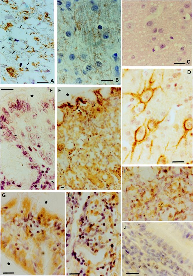
Experimental lemurs either were infected orally with the agent of bovine spongiform encephalopathy (BSE) or were maintained as uninfected control animals. Immunohistochemical examination for proteinase-resistant protein (prion protein or PrP) was performed on tissues from two infected but still asymptomatic lemurs, killed 5 months after infection, and from three uninfected control lemurs. Control tissues showed no staining, whereas PrP was detected in the infected animals in tonsil, gastrointestinal tract and associated lymphatic tissues, and spleen. In addition, PrP was detected in ventral and dorsal roots of the cervical spinal cord, and within the spinal cord PrP could be traced in nerve tracts as far as the cerebral cortex. Similar patterns of PrP immunoreactivity were seen in two symptomatic and 18 apparently healthy lemurs in three different French primate centers, all of which had been fed diets supplemented with a beef protein product manufactured by a British company that has since ceased to include beef in its veterinary nutritional products. This study of BSE-infected lemurs early in their incubation period extends previous pathogenesis studies of the distribution of infectivity and PrP in natural and experimental scrapie. The similarity of neuropathology and PrP immunostaining patterns in experimentally infected animals to those observed in both symptomatic and asymptomatic animals in primate centers suggests that BSE contamination of zoo animals may have been more widespread than is generally appreciated.
Figures
References
-
- Bons N, Mestre-Francés N, Charnay Y, Tagliavini F. Lancet. 1996;348:55. - PubMed
-
- Bons N, Mestre-Francés N, Guiraud I, Charnay Y. C R Acad Sci. 1997;320:971–979. - PubMed
-
- Tagliavini F, Prelli F, Giaccone G, Forloni G, Salmona M, Piccardo P, Ghetti B, Frangione B, Bugiani O. In: Methods in Molecular Medicine: Prion Diseases. Baker H, Ridley M, editors. Totowa, NJ: Humana; 1996. pp. 265–283.
-
- Delacourte A, Flament S, Dibe E M, Hublau P, Sablonnière B, Hemon B, Défossez A. Acta Neuropathol. 1990;80:111–117. - PubMed
-
- Bons N, Silhol S, Barbié V, Mestre-Francés N, Albe-Fessard D. Brain Res Bull. 1998;46:1–173. - PubMed
Publication types
MeSH terms
Substances
LinkOut - more resources
Full Text Sources
Medical
Research Materials
