

Herpes simplex virus vector-mediated expression of Bcl-2 prevents 6-hydroxydopamine-induced degeneration of neurons in the substantia nigra in vivo
- PMID: 10097166
- PMCID: PMC22423
- DOI: 10.1073/pnas.96.7.4078
Herpes simplex virus vector-mediated expression of Bcl-2 prevents 6-hydroxydopamine-induced degeneration of neurons in the substantia nigra in vivo
Abstract
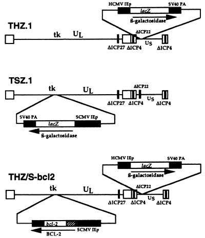
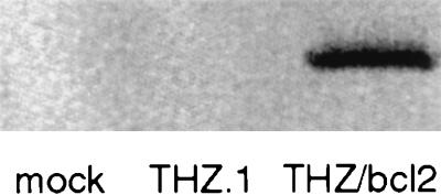
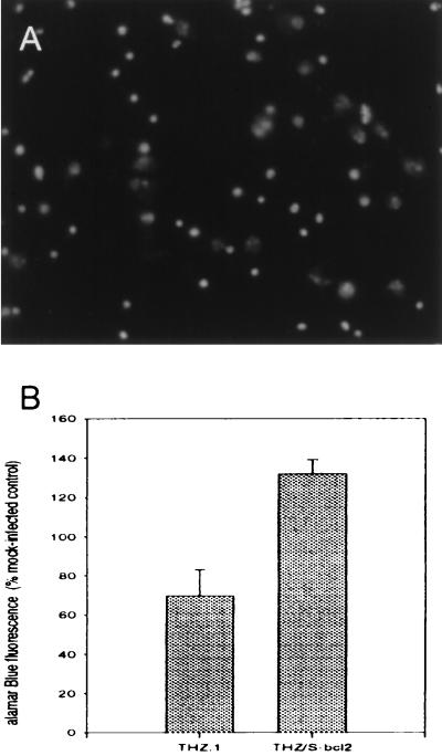
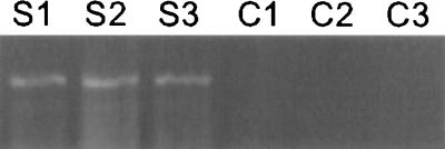
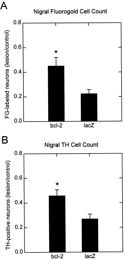
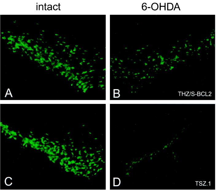
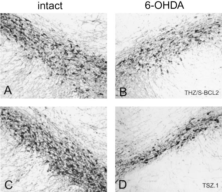
6-Hydroxydopamine (6-OHDA) is widely used to selectively lesion dopaminergic neurons of the substantia nigra (SN) in the creation of animal models of Parkinson's disease. In vitro, the death of PC-12 cells caused by exposure to 6-OHDA occurs with characteristics consistent with an apoptotic mechanism of cell death. To test the hypothesis that apoptotic pathways are involved in the death of dopaminergic neurons of the SN caused by 6-OHDA, we created a replication-defective genomic herpes simplex virus-based vector containing the coding sequence for the antiapoptotic peptide Bcl-2 under the transcriptional control of the simian cytomegalovirus immediate early promoter. Transfection of primary cortical neurons in culture with the Bcl-2-producing vector protected those cells from naturally occurring cell death over 3 weeks. Injection of the Bcl-2-expressing vector into SN of rats 1 week before injection of 6-OHDA into the ipsilateral striatum increased the survival of neurons in the SN, detected either by retrograde labeling of those cells with fluorogold or by tyrosine hydroxylase immunocytochemistry, by 50%. These results, demonstrating that death of nigral neurons induced by 6-OHDA lesioning may be blocked by the expression of Bcl-2, are consistent with the notion that cell death in this model system is at least in part apoptotic in nature and suggest that a Bcl-2-expressing vector may have therapeutic potential in the treatment of Parkinson's disease.
Figures
Similar articles
-
Bcl-2 and GDNF delivered by HSV-mediated gene transfer act additively to protect dopaminergic neurons from 6-OHDA-induced degeneration.Exp Neurol. 2001 Jun;169(2):231-8. doi: 10.1006/exnr.2001.7671. Exp Neurol. 2001. PMID: 11358438
-
Alterations in the cellular distribution of bcl-2, bcl-x and bax in the adult rat substantia nigra following striatal 6-hydroxydopamine lesions.J Neurocytol. 2004 Mar;33(2):213-23. doi: 10.1023/b:neur.0000030696.62829.ec. J Neurocytol. 2004. PMID: 15322379
-
Delivery of a GDNF gene into the substantia nigra after a progressive 6-OHDA lesion maintains functional nigrostriatal connections.Exp Neurol. 2000 Nov;166(1):1-15. doi: 10.1006/exnr.2000.7463. Exp Neurol. 2000. PMID: 11031079
-
6-Hydroxydopamine increases the level of TNFalpha and bax mRNA in the striatum and induces apoptosis of dopaminergic neurons in hemiparkinsonian rats.Brain Res. 2004 Jan 23;996(2):237-45. doi: 10.1016/j.brainres.2003.10.035. Brain Res. 2004. PMID: 14697501
-
Nigrostriatal neuronal death in Parkinson's disease--a passive or an active genetically-controlled process?J Neural Transm Suppl. 1997;49:69-76. doi: 10.1007/978-3-7091-6844-8_7. J Neural Transm Suppl. 1997. PMID: 9266415 Review.
Cited by
-
Herpes simplex virus-based vectors.Int J Exp Pathol. 2004 Oct;85(4):177-90. doi: 10.1111/j.0959-9673.2004.00383.x. Int J Exp Pathol. 2004. PMID: 15312123 Free PMC article. Review.
-
Future of cell and gene therapies for Parkinson's disease.Ann Neurol. 2008 Dec;64 Suppl 2(0 2):S122-38. doi: 10.1002/ana.21473. Ann Neurol. 2008. PMID: 19127583 Free PMC article.
-
The beneficial role of thiamine in Parkinson disease.CNS Neurosci Ther. 2013 Jul;19(7):461-8. doi: 10.1111/cns.12078. Epub 2013 Mar 6. CNS Neurosci Ther. 2013. PMID: 23462281 Free PMC article. Review.
-
Neuroprotective Effect of β-Lapachone in MPTP-Induced Parkinson's Disease Mouse Model: Involvement of Astroglial p-AMPK/Nrf2/HO-1 Signaling Pathways.Biomol Ther (Seoul). 2019 Mar 1;27(2):178-184. doi: 10.4062/biomolther.2018.234. Biomol Ther (Seoul). 2019. PMID: 30739428 Free PMC article.
-
Pseudotyping of glycoprotein D-deficient herpes simplex virus type 1 with vesicular stomatitis virus glycoprotein G enables mutant virus attachment and entry.J Virol. 2000 Mar;74(5):2481-7. doi: 10.1128/jvi.74.5.2481-2487.2000. J Virol. 2000. PMID: 10666285 Free PMC article.
References
-
- Heikkila R E, Hess A, Duvoisin R C. Science. 1984;224:1451–1453. - PubMed
-
- Nicklas W J, Vyas I, Heikkila R E. Life Sci. 1985;36:2503–2508. - PubMed
-
- Ungerstedt U. Eur J Pharmacol. 1968;5:107–110. - PubMed
-
- Ungerstedt U, Arbuthnott G W. Brain Res. 1970;24:485–493. - PubMed
-
- Jeon B S, Jackson-Lewis V, Burke R E. Neurodegeneration. 1995;4:131–137. - PubMed
Publication types
MeSH terms
Substances
LinkOut - more resources
Full Text Sources
Other Literature Sources
