Gene disruption of p27(Kip1) allows cell proliferation in the postnatal and adult organ of corti
- PMID: 10097167
- PMCID: PMC22424
- DOI: 10.1073/pnas.96.7.4084
Gene disruption of p27(Kip1) allows cell proliferation in the postnatal and adult organ of corti
Abstract
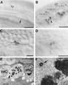
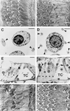
Hearing loss is most often the result of hair-cell degeneration due to genetic abnormalities or ototoxic and traumatic insults. In the postembryonic and adult mammalian auditory sensory epithelium, the organ of Corti, no hair-cell regeneration has ever been observed. However, nonmammalian hair-cell epithelia are capable of regenerating sensory hair cells as a consequence of nonsensory supporting-cell proliferation. The supporting cells of the organ of Corti are highly specialized, terminally differentiated cell types that apparently are incapable of proliferation. At the molecular level terminally differentiated cells have been shown to express high levels of cell-cycle inhibitors, in particular, cyclin-dependent kinase inhibitors [Parker, S. B., et al. (1995) Science 267, 1024-1027], which are thought to be responsible for preventing these cells from reentering the cell cycle. Here we report that the cyclin-dependent kinase inhibitor p27(Kip1) is selectively expressed in the supporting-cell population of the organ of Corti. Effects of p27(Kip1)-gene disruption include ongoing cell proliferation in postnatal and adult mouse organ of Corti at time points well after mitosis normally has ceased during embryonic development. This suggests that release from p27(Kip1)-induced cell-cycle arrest is sufficient to allow supporting-cell proliferation to occur. This finding may provide an important pathway for inducing hair-cell regeneration in the mammalian hearing organ.
Figures


Similar articles
-
p27(Kip1) links cell proliferation to morphogenesis in the developing organ of Corti.Development. 1999 Apr;126(8):1581-90. doi: 10.1242/dev.126.8.1581. Development. 1999. PMID: 10079221
-
The Hippo pathway and p27Kip1 cooperate to suppress mitotic regeneration in the organ of Corti and the retina.Proc Natl Acad Sci U S A. 2025 Apr 8;122(14):e2411313122. doi: 10.1073/pnas.2411313122. Epub 2025 Apr 3. Proc Natl Acad Sci U S A. 2025. PMID: 40178894
-
p27(Kip1) deficiency causes organ of Corti pathology and hearing loss.Hear Res. 2006 Apr;214(1-2):28-36. doi: 10.1016/j.heares.2006.01.014. Epub 2006 Mar 2. Hear Res. 2006. PMID: 16513305
-
Postnatal development, maturation and aging in the mouse cochlea and their effects on hair cell regeneration.Hear Res. 2013 Mar;297:68-83. doi: 10.1016/j.heares.2012.11.009. Epub 2012 Nov 16. Hear Res. 2013. PMID: 23164734 Free PMC article. Review.
-
Multiple functions of p27(Kip1) and its alterations in tumor cells: a review.J Cell Physiol. 2000 Apr;183(1):18-27. doi: 10.1002/(SICI)1097-4652(200004)183:1<18::AID-JCP3>3.0.CO;2-S. J Cell Physiol. 2000. PMID: 10699962 Review.
Cited by
-
Decellularized ear tissues as scaffolds for stem cell differentiation.J Assoc Res Otolaryngol. 2013 Feb;14(1):3-15. doi: 10.1007/s10162-012-0355-y. Epub 2012 Oct 20. J Assoc Res Otolaryngol. 2013. PMID: 23085833 Free PMC article.
-
LGR4 and LGR5 Regulate Hair Cell Differentiation in the Sensory Epithelium of the Developing Mouse Cochlea.Front Cell Neurosci. 2016 Aug 5;10:186. doi: 10.3389/fncel.2016.00186. eCollection 2016. Front Cell Neurosci. 2016. PMID: 27559308 Free PMC article.
-
Sensory hair cell death and regeneration in fishes.Front Cell Neurosci. 2015 Apr 21;9:131. doi: 10.3389/fncel.2015.00131. eCollection 2015. Front Cell Neurosci. 2015. PMID: 25954154 Free PMC article. Review.
-
Where hearing starts: the development of the mammalian cochlea.J Anat. 2016 Feb;228(2):233-54. doi: 10.1111/joa.12314. Epub 2015 Jun 5. J Anat. 2016. PMID: 26052920 Free PMC article. Review.
-
Generation of inner ear hair cells by direct lineage conversion of primary somatic cells.Elife. 2020 Jun 30;9:e55249. doi: 10.7554/eLife.55249. Elife. 2020. PMID: 32602462 Free PMC article.
References
Publication types
MeSH terms
Substances
Grants and funding
LinkOut - more resources
Full Text Sources
Other Literature Sources
Molecular Biology Databases
Miscellaneous

