

Coordinate regulation of gonadotropin-releasing hormone neuronal firing patterns by cytosolic calcium and store depletion
- PMID: 10097170
- PMCID: PMC22427
- DOI: 10.1073/pnas.96.7.4101
Coordinate regulation of gonadotropin-releasing hormone neuronal firing patterns by cytosolic calcium and store depletion
Abstract
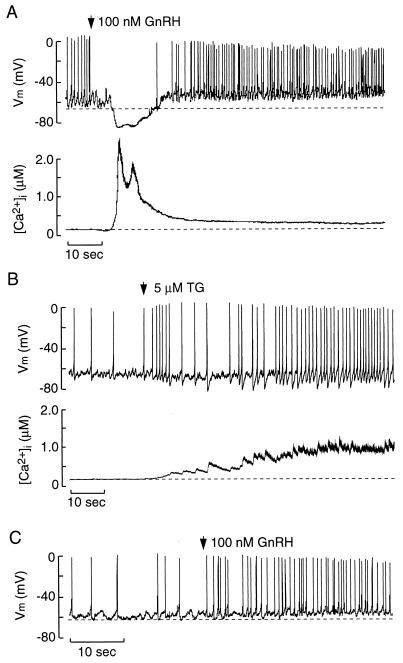
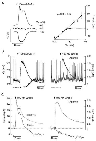
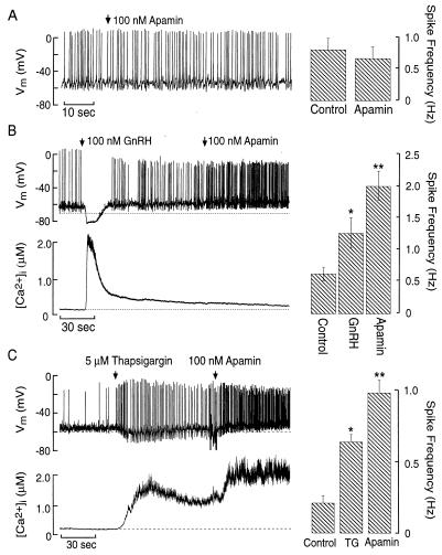
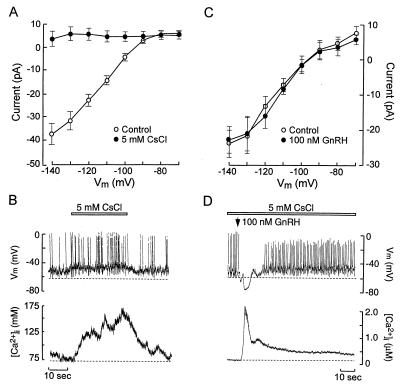
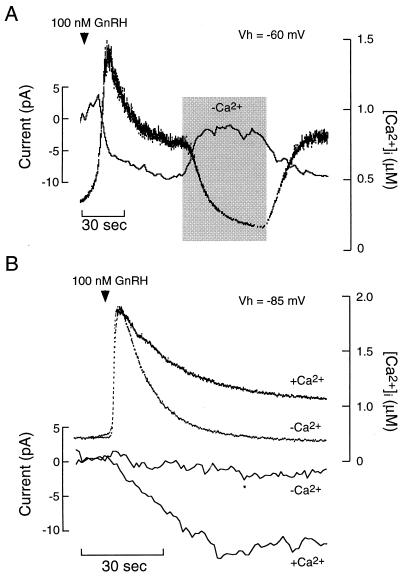
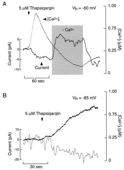
Elevation of cytosolic free Ca2+ concentration ([Ca2+]i) in excitable cells often acts as a negative feedback signal on firing of action potentials and the associated voltage-gated Ca2+ influx. Increased [Ca2+]i stimulates Ca2+-sensitive K+ channels (IK-Ca), and this, in turn, hyperpolarizes the cell and inhibits Ca2+ influx. However, in some cells expressing IK-Ca the elevation in [Ca2+]i by depletion of intracellular stores facilitates voltage-gated Ca2+ influx. This phenomenon was studied in hypothalamic GT1 neuronal cells during store depletion caused by activation of gonadotropin-releasing hormone (GnRH) receptors and inhibition of endoplasmic reticulum (Ca2+)ATPase with thapsigargin. GnRH induced a rapid spike increase in [Ca2+]i accompanied by transient hyperpolarization, followed by a sustained [Ca2+]i plateau during which the depolarized cells fired with higher frequency. The transient hyperpolarization was caused by the initial spike in [Ca2+]i and was mediated by apamin-sensitive IK-Ca channels, which also were operative during the subsequent depolarization phase. Agonist-induced depolarization and increased firing were independent of [Ca2+]i and were not mediated by inhibition of K+ current, but by facilitation of a voltage-insensitive, Ca2+-conducting inward current. Store depletion by thapsigargin also activated this inward depolarizing current and increased the firing frequency. Thus, the pattern of firing in GT1 neurons is regulated coordinately by apamin-sensitive SK current and store depletion-activated Ca2+ current. This dual control of pacemaker activity facilitates voltage-gated Ca2+ influx at elevated [Ca2+]i levels, but also protects cells from Ca2+ overload. This process may also provide a general mechanism for the integration of voltage-gated Ca2+ influx into receptor-controlled Ca2+ mobilization.
Figures
Similar articles
-
Autocrine regulation of calcium influx and gonadotropin-releasing hormone secretion in hypothalamic neurons.Biochem Cell Biol. 2000;78(3):359-70. Biochem Cell Biol. 2000. PMID: 10949086 Review.
-
Ca2+ entry in gonadotrophs and alpha T3-1 cells: does store-dependent Ca2+ influx mediate gonadotrophin-releasing hormone action?J Endocrinol. 1996 Apr;149(1):155-69. doi: 10.1677/joe.0.1490155. J Endocrinol. 1996. PMID: 8676048
-
Control of action potential-driven calcium influx in GT1 neurons by the activation status of sodium and calcium channels.Mol Endocrinol. 1999 Apr;13(4):587-603. doi: 10.1210/mend.13.4.0261. Mol Endocrinol. 1999. PMID: 10194765
-
Gonadotropin-releasing hormone-induced calcium signaling in clonal pituitary gonadotrophs.Endocrinology. 1992 Aug;131(2):925-32. doi: 10.1210/endo.131.2.1379169. Endocrinology. 1992. PMID: 1379169
-
Ion Channels of Pituitary Gonadotrophs and Their Roles in Signaling and Secretion.Front Endocrinol (Lausanne). 2017 Jun 9;8:126. doi: 10.3389/fendo.2017.00126. eCollection 2017. Front Endocrinol (Lausanne). 2017. PMID: 28649232 Free PMC article. Review.
Cited by
-
Modeling of membrane excitability in gonadotropin-releasing hormone-secreting hypothalamic neurons regulated by Ca2+-mobilizing and adenylyl cyclase-coupled receptors.J Neurosci. 2000 Dec 15;20(24):9290-7. doi: 10.1523/JNEUROSCI.20-24-09290.2000. J Neurosci. 2000. PMID: 11125008 Free PMC article.
-
Amplitude-dependent spike-broadening and enhanced Ca(2+) signaling in GnRH-secreting neurons.Biophys J. 2000 Sep;79(3):1310-23. doi: 10.1016/S0006-3495(00)76384-3. Biophys J. 2000. PMID: 10968994 Free PMC article.
-
Differential regulation of GnRH secretion in the preoptic area (POA) and the median eminence (ME) in male mice.Endocrinology. 2015 Jan;156(1):231-41. doi: 10.1210/en.2014-1458. Endocrinology. 2015. PMID: 25314270 Free PMC article.
-
Cell type-specific expression of a genetically encoded calcium indicator reveals intrinsic calcium oscillations in adult gonadotropin-releasing hormone neurons.J Neurosci. 2007 Jan 24;27(4):860-7. doi: 10.1523/JNEUROSCI.3579-06.2007. J Neurosci. 2007. PMID: 17251427 Free PMC article.
-
Regulation of reproduction via tight control of gonadotropin hormone levels.Mol Cell Endocrinol. 2018 Mar 5;463:116-130. doi: 10.1016/j.mce.2017.03.022. Epub 2017 Mar 22. Mol Cell Endocrinol. 2018. PMID: 28342855 Free PMC article. Review.
References
MeSH terms
Substances
LinkOut - more resources
Full Text Sources
Miscellaneous
