

Overexpression of thioredoxin in transgenic mice attenuates focal ischemic brain damage
- PMID: 10097175
- PMCID: PMC22432
- DOI: 10.1073/pnas.96.7.4131
Overexpression of thioredoxin in transgenic mice attenuates focal ischemic brain damage
Abstract
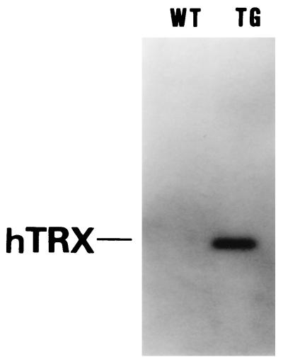
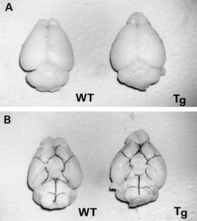
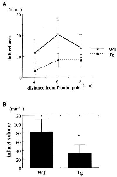
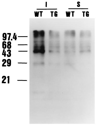
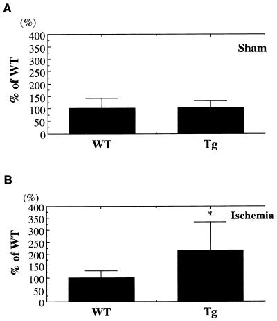
Thioredoxin (TRX) plays important biological roles both in intra- and extracellular compartments, including in regulation of various intracellular molecules via thiol redox control. We produced TRX overexpressing mice and confirmed that there were no anatomical and physiological differences between wild-type (WT) mice and TRX transgenic (Tg) mice. In the present study we subjected mice to focal brain ischemia to shed light on the role of TRX in brain ischemic injury. At 24 hr after middle cerebral artery occlusion, infarct areas and volume were significantly smaller in Tg mice than in WT mice. Moreover neurological deficit was ameliorated in Tg mice compared with WT mice. Protein carbonyl content, a marker of cellular protein oxidation, in Tg mice showed less increase than did that of WT mice after the ischemic insult. Furthermore, c-fos expression in Tg mice was stronger than in WT mice 1 hr after ischemia. Our results suggest that transgene expression of TRX decreased ischemic neuronal injury and that TRX and the redox state modified by TRX play a crucial role in brain damage during stroke.
Figures

Similar articles
-
Mice overexpressing extracellular superoxide dismutase have increased resistance to focal cerebral ischemia.Neuroscience. 1999 Jan;88(1):185-91. doi: 10.1016/s0306-4522(98)00208-5. Neuroscience. 1999. PMID: 10051199
-
Human copper-zinc superoxide dismutase transgenic mice are highly resistant to reperfusion injury after focal cerebral ischemia.Stroke. 1994 Jan;25(1):165-70. doi: 10.1161/01.str.25.1.165. Stroke. 1994. PMID: 8266365
-
Attenuation of neuronal degeneration in thioredoxin-1 overexpressing mice after mild focal ischemia.Brain Res. 2009 May 26;1272:62-70. doi: 10.1016/j.brainres.2009.03.023. Epub 2009 Mar 25. Brain Res. 2009. PMID: 19328186
-
Thioredoxin as a Therapeutic Target in Cerebral Ischemia.Curr Pharm Des. 2018;24(25):2986-2992. doi: 10.2174/1381612824666180820143853. Curr Pharm Des. 2018. PMID: 30124144 Review.
-
Role of superoxide dismutase in ischemic brain injury: a study using SOD-1 transgenic mice.Cell Mol Neurobiol. 1998 Dec;18(6):609-20. doi: 10.1023/a:1020677701368. Cell Mol Neurobiol. 1998. PMID: 9876869 Free PMC article. Review.
Cited by
-
Induction of antioxidative and antiapoptotic thioredoxin supports neuroprotective hypothesis of estrogen.Endocrine. 2003 Jun;21(1):27-31. doi: 10.1385/endo:21:1:27. Endocrine. 2003. PMID: 12777700 Review.
-
Stroke Proteomics: From Discovery to Diagnostic and Therapeutic Applications.Circ Res. 2022 Apr 15;130(8):1145-1166. doi: 10.1161/CIRCRESAHA.122.320110. Epub 2022 Apr 14. Circ Res. 2022. PMID: 35420912 Free PMC article. Review.
-
Delayed administration of alpha-difluoromethylornithine prevents hippocampus-dependent cognitive impairment after single and combined injury in mice.Radiat Res. 2014 Nov;182(5):489-98. doi: 10.1667/RR13753.1. Epub 2014 Nov 6. Radiat Res. 2014. PMID: 25375198 Free PMC article.
-
Redox compartmentalization in eukaryotic cells.Biochim Biophys Acta. 2008 Nov;1780(11):1273-90. doi: 10.1016/j.bbagen.2008.01.011. Epub 2008 Jan 26. Biochim Biophys Acta. 2008. PMID: 18267127 Free PMC article. Review.
-
Oxidative stress and diabetes: what can we learn about insulin resistance from antioxidant mutant mouse models?Free Radic Biol Med. 2012 Jan 1;52(1):46-58. doi: 10.1016/j.freeradbiomed.2011.10.441. Epub 2011 Oct 20. Free Radic Biol Med. 2012. PMID: 22056908 Free PMC article. Review.
References
-
- Siesjo B K. J Neurosurg. 1984;60:883–908. - PubMed
-
- Chan P H. Brain Pathol. 1994;4:59–65. - PubMed
-
- Choi D W. Neuron. 1988;1:623–634. - PubMed
-
- Eliasson M J L, Sampei K, Mandir A S, Hurn P D, Traystman R J, Bao J, Pieper A, Wang Z O, Dawson T M, Snyder S H, Dawson V L. Nat Med. 1997;3:1089–1095. - PubMed
Publication types
MeSH terms
Substances
LinkOut - more resources
Full Text Sources
Other Literature Sources
Medical
Molecular Biology Databases
Miscellaneous
