

Review
doi: 10.1016/s0167-5699(98)01398-x.
CD13--not just a marker in leukemia typing
Affiliations
- PMID: 10098327
- PMCID: PMC7141563
- DOI: 10.1016/s0167-5699(98)01398-x
Item in Clipboard
Review
CD13--not just a marker in leukemia typing
Immunol Today.
1999 Feb.
Abstract
Membrane peptidases are a multifunctional group of ectoenzymes that have been implicated in the control of growth and differentiation of many cellular systems. Here, using aminopeptidase N/CD13 as an example, Dagmar Riemann and colleagues discuss the role of cell-cell contact in peptidase regulation and the influence of peptidases on cellular functions.
Figures
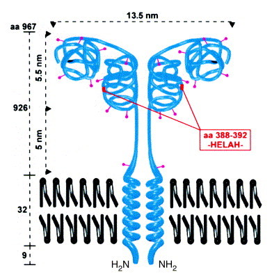
Structure of CD13. The type II membrane protein is found as a dimer of two non-covalently associated subunits with a relative molecular mass of 160 kDa each. CD13 has ten N-glycosylation sites (∼400 carbohydrate residues comprising approximately 40% of the molecular mass). Analogous to aminopeptidase A (APA), a two-domain structure can be hypothesized with a substrate-binding site in the C-terminal and the active center in the N-terminal domain. The dimensions of the molecule are taken from Noren et al.
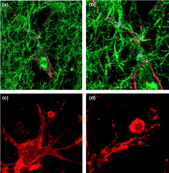
Confocal laser scanning micrographs of CD13 surface expression on fibroblast-like synoviocytes and on lymphocytes in a three-dimensional collagen matrix. (a) Confocal reflection contrast in combination with immunofluorescence staining is used to show surface expression of CD13 on synoviocytes (red) as well as reflection of collagen (type I) fibers (green). (b) Part of (a) in higher magnification. Points of close contact between collagen fibers (reflection mode) and CD13 surface staining on the synoviocyte membrane (immunofluorescence) appear in blue. (c) and (d) Immunofluorescence localization of CD13 on synoviocytes and on T cells. Peripheral blood lymphocytes were allowed to migrate into a three-dimensional collagen lattice and to adhere to synoviocytes within 3 h at 37°C. Cells were subsequently stained for CD13 (red) and fixed. Magnification for (a), ×1000; (b), ×1700; (c), ×1000; (d), ×1900.
References
Publication types
MeSH terms
Substances
LinkOut - more resources
Full Text Sources
Other Literature Sources
Miscellaneous
