. 1999 Feb;78(2):117-26.
doi: 10.1016/S0171-9335(99)80013-2.

2E4 (kaptin): a novel actin-associated protein from human blood platelets found in lamellipodia and the tips of the stereocilia of the inner ear

Affiliations

2E4 (kaptin): a novel actin-associated protein from human blood platelets found in lamellipodia and the tips of the stereocilia of the inner ear

E L Bearer et al. Eur J Cell Biol. 1999 Feb.

Abstract

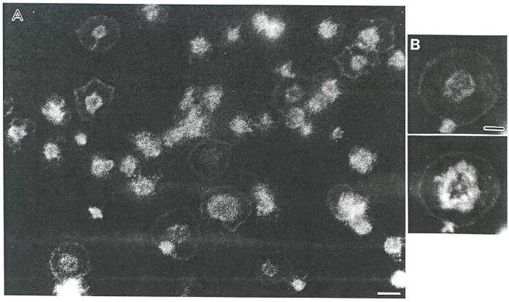
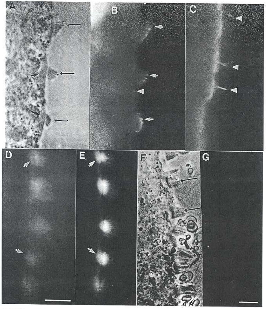
Platelet activation, crucial for hemostasis, requires actin polymerization, yet the molecular mechanisms by which localized actin polymerization is mediated are not clear. Here we report the characterization of a novel actin-binding protein, 2E4, originally isolated from human blood platelets and likely to be involved in the actin rearrangements occurring during activation. 2E4 binds to filamentous (F)-actin by F-actin affinity chromatography and is eluted from F-actin affinity columns and extracted from cells with ATP. Its presence at the leading edge of platelets spread on glass and in the lamellipodia of motile fibroblasts suggests a role in actin dynamics. Using localization to obtain clues about function, we stained the sensory epithelium of the embryonic inner ear to determine whether 2E4 is at the barbed end of actin filaments during their elongation. Indeed, 2E4 was present at the tips of the elongating stereocilium. 2E4 is novel by DNA sequence and has no identifiable structural motifs. Its unusual amino acid sequence, its ATP-sensitive actin association and its location at sites of actin polymerization in cells suggest 2E4 plays a unique role in the actin rearrangements that accompany platelet activation and stereocilia formation.


Figures

Fig. 1
Fig. 1
2E4 elutes from F-actin affinity columns with ATP. A. Coomassie-stained 6–12% gradient SDS-gel of H, ADP-activated platelet homogenate; X, extract of same preparation after clarifying centrifugation which is loaded in parallel onto two columns, one cross-linked with F-actin and the other with albumin (BSA); W, last wash before elution from either column; ATP, fraction containing the peak protein eluted with 5 mM ATP from either column; KC1, peak protein fraction eluted with 1 M KCl from either column. Bars indicate molecular weight standards 200, 98, 68, 45, and 31 kDa. B. Corresponding Western blot of the same samples probed with MAb 2E4. Order of lanes is the same as in (A). Bar indicates 45 kDa standard. Note that a ~45 kDa band appears in homogenate, extract and the ATP elution from the F-actin column only.
Fig. 2
Fig. 2
2E4 is localized in the lamellipodia of platelets spread on glass. A. Low magnification of a field of platelets spread for 15 min on glass and stained with MAb 2E4, Bar = 5 µm. B. An individual platelet shown at higher magnification double-stained for 2E4 (upper) and F-actin with phalloidin (lower). Note that the staining coincides at the periphery but not in the center of the cell. Bar = 2µm.
Fig. 3
Fig. 3
MAb 2E4 recognizes ~45 kDa protein in Western blots of other cells with specialized actin structures. Upper panel: Coomassie-stained 10% SDS-gel of specimen as indicated. Molecular weight standards at the left; 200, 98, 45, 31 kDa. Platelets are human and all other samples are chick. Lower panel: Corresponding Western blot of the same samples probed with MAb 2E4. Bands identified by the antibody are the same size, ~45 kDa, in all lanes. In the platelet sample, there is also a faint 55 kDa band as well as some staining at the dye front. Note that the antibody recognizes a single 45 kDa band in chick samples.
Fig. 4
Fig. 4
2E4 is located in the lamellipodia of chick embryo fibroblasts, Double-labeled images of chick embryo fibroblasts stained with MAb 2E4 (A, C) and for actin with phalloidin (B, D). Arrows in A and B indicate adhesion plaques, visible when stained for actin but not stained by 2E4. Bar = 5µm.
Fig. 5
Fig. 5
2E4 is located at the distal tips of stereocilia of hair cells in the inner ear. A. Phase-contrast image of sensory epithelium from the chick cochlea. Long arrows indicate staircase of stereocilia projecting from the apical surface of the hair cells. Short arrows indicate position of tectal plate. B. Same field as in (A) imaged by fluorescence microscopy to show 2E4 staining, which is limited to the outer 1/3 of the stereocilia (arrows). Note possible weak staining of short microvilli on support cell (arrowhead) and no staining of tectal plate. C. A different sensory epithelium stained for microtubules, Arrowheads indicate the primary cilium, a microtubule-rich structure. D. Another specimen, stained with an IgM anti-actin monoclonal antibody. Note that the entire stereocilium is stained, as well as the tectal plate in the apical cytoplasm of the hair cell. Bar = 10 µm. E. Another specimen labeled with phalloidin for F-actin. Note that the distribution of staining matches that of anti-actin. F. Phase-contrast image of a cochlear sensory epithelium that was extracted with 5 mM ATP and detergent before fixation. Note that the stereocilia are intact (arrows) although there is substantial membranous debris. G. Same fields as (F), imaged with fluorescence for 2E4. No staining is detected. Bar shown in (G) indicates magnification for AC, EG and corresponds to 10 µm.
Fig. 6
Fig. 6
Semi-purification of 2E4 by anion exchange chromatography. A. Coomassie-stained gel of peak fractions from a DEAE column loaded with activated platelet extract and eluted with a 0.02–1.0 M salt gradient. Note that each fraction is enriched with a different set of protein species. Molecular weights: 200, 98, 68, 45, 31 kDa. B. Corresponding Western blot loaded with the same samples as (A) and probed with MAb 2E4. Arrows in A and B indicate 45 kDa. Note the ~45 kDa band in fraction 7 in B. Molecular weight standards: 200, 98, 68, 45 kDa.
Fig. 7
Fig. 7
Amino acid sequence of 2E4. Peptide sequences obtained from the 45 kDa band recognized by MAb 2E4 in fraction 7 of anion exchange separation of platelet proteins are underlined. No motifs or homologous proteins were found in the data bank, although ESTs that matched the DNA sequence were found in both mouse and human libraries. Accession number of 2E4; (GenBank AF105369).
Fig. 8
Fig. 8
Monoclonal antibody recognizes bacterially expressed 2E4 protein, and polyclonal antibodies recognize a 45 kDa band in platelet homogenates. Coomassie-stained 12% SDS gel (Gel) loaded with bacterially expressed full-length 2E4 protein purified by nickel column (2E4) and bacterially expressed full-length Arp2 also isolated by nickel column (Arp2), 45 kDa band indicated by arrow at left. A parallel blot probed with MAb 2E4 (MAb Blot) shows that MAb 2E4 recognizes the 2E4 protein (2E4) but not Arp 2 (Arp 2) pAb shows a Coomassie-stained 8.5% SDS gel of platelet homogenates (gel) and corresponding Western blot (blot) probed with affinity-purified polyclonal anti-bodies raised against bacterially expressed cloned 2E4. Molecular weight markers in pAb indicated to the left: 200, 98, 68, 45, 31 kDa.
Fig. 9
Fig. 9
Polyclonal antibodies raised against the cloned protein stain the lamellipodia of spreading platelets. A. Platelets spread on glass stained with polyclonal anti-2E4. B. Same two platelets, double-labeled with phalloidin, Bar = 2.5 µm.

References

    1. Albanesi JP, Lynch TJ, Fujosaki H, Bowerts B, Korn ED. Purification and characterization of an ATP-sensitive actin gelation factor protein from Acanthamoeba castellani. J. Biol. Chem. 1987;262:3404–3408. - PubMed
    1. Aroian RV, Field C, Pruliere G, Kenyon C, Alberts BM. Isolation of actin-associated proteins from Caenorhabditis elegans oocytes and their localization in the early embryo. EMBO J. 1997;16:1541–1549. - PMC - PubMed
    1. Bearer EL. Platelet membrane skeleton revealed by quick freeze-deep etch. Anat. Rec. 1990;227:1–11. - PMC - PubMed
    1. Bearer EL. Direct observation of actin filament severing by gelsolin and binding by gCap 39 and CapZ. J. Cell Biol. 1991;115:1629–1638. - PMC - PubMed
    1. Bearer EL. An actin-associated protein present in the microtubule organizing center and the growth cones of PC-12 cells. J. Neurosci. 1992;12:750–761. - PMC - PubMed

MeSH terms

Associated data