

doi: 10.1126/science.284.5411.92.
Complexity in biological signaling systems
Affiliations
- PMID: 10102825
- PMCID: PMC3773983
- DOI: 10.1126/science.284.5411.92
Item in Clipboard
Complexity in biological signaling systems
Science.
.
Abstract
Biological signaling pathways interact with one another to form complex networks. Complexity arises from the large number of components, many with isoforms that have partially overlapping functions; from the connections among components; and from the spatial relationship between components. The origins of the complex behavior of signaling networks and analytical approaches to deal with the emergent complexity are discussed here.
Figures
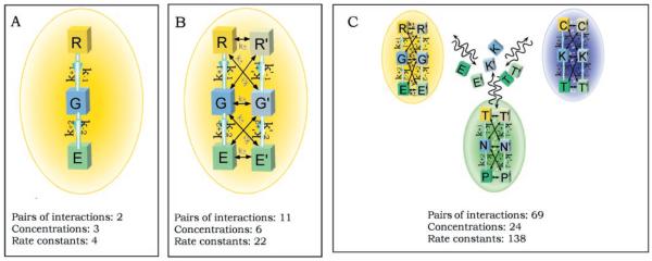
The increasing complexity of signaling pathways inside a cell. In each panel, k is the rate constant for the first pathway and k′ represents constants for the second pathway; plus and minus signs indicate forward and reverse, respectively; 1 and 2 indicate pathways 1 and 2. (A) A simple three-component pathway. The arrows indicate the direction of the signal flow. Each component interacts only with its adjacent component. This system represents a typical design of a transmembrane G protein signaling pathway, and the lettering for the components R (receptor), G (G protein), and E (effector) reflects this. (B) Two interactive signaling pathways in one compartment. Here, interactions are restricted to adjacent partners to represent real situations and limit the complexity of the system. (C) A complex system consisting of two interactive-pathways in each of three interacting compartments, colored yellow, blue, and green. Such a system could represent the first level of compartmentalization of the cell into membrane, cytoplasmic, and nuclear compartments. C, cytoplasmic components; K, kinase; T, transcription factors; N, nucleic acids; P, protein components in the nucleus. The communications between the compartments are carried out by the translocation of the signaling molecules. The number of interactions and the minimal number of parameters (concentration of reactants plus rate constants) for each system is given. The increasing complexity in terms of the number of parameters needed to specify the system can be readily seen.
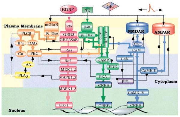
Four major signaling pathways in the postsynaptic region of a neuron that combine to form a local signaling network. The major linear routes of signal flow are depicted by the thick arrows of four different colors: orange [phospholipase C (PLC) pathway], pink (Ras pathway), green (adenylyl cyclase pathway), and blue [Ca2+/calmodulin (CaM) pathway]. The interactions between different pathways are represented by black lines with arrow (representing activation) or a dot (representing inhibition). Although most major interactions in the network are shown, these connections are not meant to be all-inclusive; additional connections could exist. The three-colored background represents three different cell compartments: the plasma membrane (light yellow), cytosol (light blue), and nucleus (light green). Some of the signaling proteins that translocate between different compartments are shown in both compartments. Examples include MAP kinase, which when activated translocates from the cytoplasm to the nucleus to phosphorylate and activate transcription factors; and the transcription factor CREB, which upon phosphorylation by protein kinase A (PKA) translocates to the nucleus.
References
-
- Alex LA, Simon MI. Trends Genet. 1994;10:133. - PubMed
-
- Segel IH. Enzyme Kinetics–Behavior and Analysis of Rapid Equilibrium and Steady State Enzyme Systems. Wiley; New York: 1975.
Publication types
MeSH terms
Substances
Grants and funding
LinkOut - more resources
Full Text Sources
Other Literature Sources
