. 1999 Apr;65(4):1644-51.
doi: 10.1128/AEM.65.4.1644-1651.1999.

Modes of action of acarbose hydrolysis and transglycosylation catalyzed by a thermostable maltogenic amylase, the gene for which was cloned from a Thermus strain

Affiliations

Modes of action of acarbose hydrolysis and transglycosylation catalyzed by a thermostable maltogenic amylase, the gene for which was cloned from a Thermus strain

T J Kim et al. Appl Environ Microbiol. 1999 Apr.

Abstract

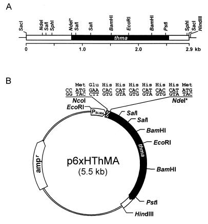
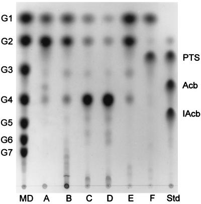
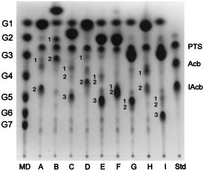
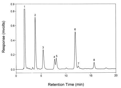
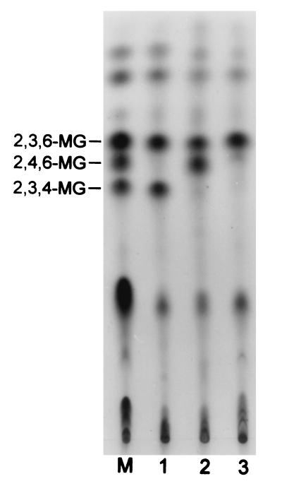
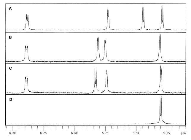
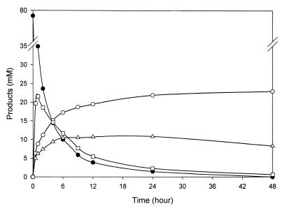
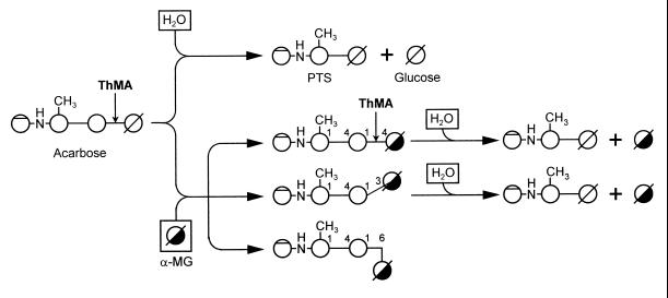
A maltogenic amylase gene was cloned in Escherichia coli from a gram-negative thermophilic bacterium, Thermus strain IM6501. The gene encoded an enzyme (ThMA) with a molecular mass of 68 kDa which was expressed by the expression vector p6xHis119. The optimal temperature of ThMA was 60 degrees C, which was higher than those of other maltogenic amylases reported so far. Thermal inactivation kinetic analysis of ThMA indicated that it was stabilized in the presence of 10 mM EDTA. ThMA harbored both hydrolysis and transglycosylation activities. It hydrolyzed beta-cyclodextrin and starch mainly to maltose and pullulan to panose. ThMA not only hydrolyzed acarbose, an amylase inhibitor, to glucose and pseudotrisaccharide (PTS) but also transferred PTS to 17 sugar acceptors, including glucose, fructose, maltose, cellobiose, etc. Structural analysis of acarbose transfer products by using methylation, thin-layer chromatography, high-performance ion chromatography, and nuclear magnetic resonance indicated that PTS was transferred primarily to the C-6 of the acceptors and at lower degrees to the C-3 and/or C-4. The transglycosylation of sugar to methyl-alpha-D-glucopyranoside by forming an alpha-(1,3)-glycosidic linkage was demonstrated for the first time by using acarbose and ThMA. Kinetic analysis of the acarbose transfer products showed that the C-4 transfer product formed most rapidly but readily hydrolyzed, while the C-6 transfer product was stable and accumulated in the reaction mixture as the main product.


Figures

FIG. 1
FIG. 1
Restriction maps of the ThMA gene clones. (A) The box represents the 2.9-kb SacI DNA fragment cloned in pThMA119. The black box indicates the location of the ThMA structural gene, directing from left to right, and the lines represent the vector. (B) For overexpression and easy purification of ThMA, the structural gene on the NdeI*-HindIII DNA fragment was fused to six histidines in frame under the control of the BLMA promoter (PBLMA) by subcloning it into p6xHis119 at the NdeI and HindIII sites. The NdeI* site was introduced into the ThMA gene for convenience in subcloning.
FIG. 2
FIG. 2
SDS-PAGE of purified ThMA. The apparent molecular mass of ThMA purified by a traditional chromatographic procedure (panel A, lane 1) and that of six-His-ThMA purified by using an Ni-NTA column (panel B, lane 2) from the E. coli lysate (panel B, lane 1) were approximately 64,000 Da. Standard size markers (lanes M) were myosin (205 kDa), β-galactosidase (116 kDa), phosphorylase b (97.4 kDa), bovine serum albumin (66 kDa), ovalbumin (45 kDa), and carbonic anhydrase (29 kDa).
FIG. 3
FIG. 3
TLC analysis of ThMA hydrolysis activity. ThMA was reacted with various substrates (0.5% [wt/vol]), including β-cyclodextrin (lane A), soluble starch (lane B), pullulan (lane C), panose (lane D), maltotriose (lane E), and acarbose (lane F). The reactions were carried out at 60°C for 12 h. Maltodextrins (MD) of G1 to G7, PTS, acarbose (Acb), and isoacarbose (IAcb) were used as standards (Std).
FIG. 4
FIG. 4
TLC analysis of acarbose transfer products. Purified ThMA transferred PTS resulting from the hydrolysis of acarbose to various acceptors: glucose (A), α-MG (B), galactose (C), fructose (D), maltose (E), cellobiose (F), lactose (G), sucrose (H), and gentiobiose (I). Maltodextrins (MD) G1 to G7, PTS, acarbose (Acb), and isoacarbose (IAcb) were used as standards (Std). The resulting transfer products were numbered (1, 2, and 3) in the order of traveling distance.
FIG. 5
FIG. 5
Results from HPIC of acarbose transfer products. Acarbose and α-MG were reacted in the presence of ThMA, and the resulting reaction mixture was subjected to HPIC. Peak 1 represents α-MG; peak 2, glucose; peak 3, α-(1,6)-linked transfer product; peak 4, α-(1,4)-linked transfer product; peak 5, α-(1,3)-linked transfer product; peak 6, PTS; peak 7, isoacarbose; and peak 8, acarbose.
FIG. 6
FIG. 6
Methylation analysis of acarbose transfer products formed with α-MG. Lane M was loaded with a mixture of methylated alternan and β-cyclodextrin. Lane 1, methylated transfer product of peak 3 in Fig. 5; lane 2, methylated transfer product of peak 5 in Fig. 5; lane 3, methylated transfer product of peak 4 in Fig. 5.
FIG. 7
FIG. 7
1H-NMR spectroscopy of α-(1,6)-acarbose transfer product (A), α-(1,4)-acarbose transfer product (B), α-(1,3)-acarbose transfer product (C), and α-MG (D). Peaks at 5.45, 5.82, and 5.85 ppm were assigned to H1 of α-(1,6)-, α-(1,4)-, and α-(1,3)-linked units between glucose and α-MG, respectively.
FIG. 8
FIG. 8
Time course analysis of acarbose transfer product formation. ThMA was reacted with acarbose (5% [wt/vol]) and α-MG (10% [wt/vol]) at 60°C for 48 h. The graph shows the hydrolysis of acarbose (black circles), formation of α-(1,4)-acarbose transfer product (squares), formation of α-(1,6)-acarbose transfer product (white circles), and formation of α-(1,3)-acarbose transfer product (triangles).
FIG. 9
FIG. 9
Proposed action modes of ThMA in the transfer reaction with acarbose and water or methyl-α-d-glucopyranoside as acceptors. If water acts as an acceptor, acarbose is hydrolyzed to PTS and glucose. If methyl-α-d-glucopyranoside (α-MG) acts as an acceptor, α-(1,4)-, α-(1,3)-, and α-(1,6)-acarbose transfer products are formed. α-(1,4)- and α-(1,3)-acarbose transfer products are hydrolyzed further to PTS and α-MG, while α-(1,6)-acarbose transfer products are accumulated without further hydrolysis.

References

    1. Aleshin A E, Firsov L M, Honzatke R B. Refined structure for the complex of acarbose with glucoamylase from Aspergillus awamori var. X100 to 2.4-Å resolution. J Biol Chem. 1994;269:15631–15639. - PubMed
    1. BMDP statistical software. 1981. California State University.
    1. Bradford M. A rapid and sensitive method for the quantitation of microgram quantities of protein utilizing the principle of protein-dye binding. Anal Biochem. 1976;72:248–254. - PubMed
    1. Brzozowski A M, Davies M J. Structure of the Aspergillus oryzae α-amylase complexed with the inhibitor acarbose at 2.0 Å. Biochemistry. 1997;36:10837–10845. - PubMed
    1. Cha H J, Yoon H G, Kim Y W, Lee H S, Kim J W, Kweon K S, Oh B H, Park K H. Molecular and enzymatic characterization of novel maltogenic amylase that hydrolyzes and transglycosylates acarbose. Eur J Biochem. 1998;253:251–262. - PubMed

Publication types

Associated data

LinkOut - more resources