. 1999 Apr 5;189(7):1053-62.
doi: 10.1084/jem.189.7.1053.

In autoimmune diabetes the transition from benign to pernicious insulitis requires an islet cell response to tumor necrosis factor alpha

Affiliations

In autoimmune diabetes the transition from benign to pernicious insulitis requires an islet cell response to tumor necrosis factor alpha

S V Pakala et al. J Exp Med. .

Abstract

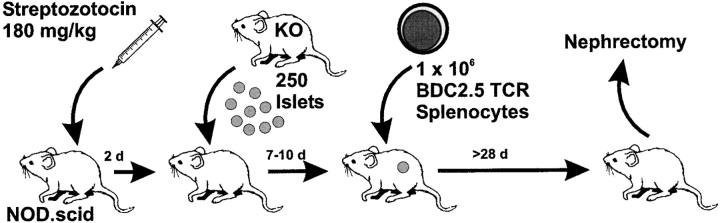
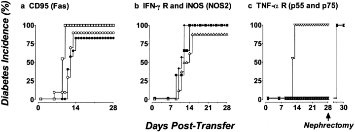
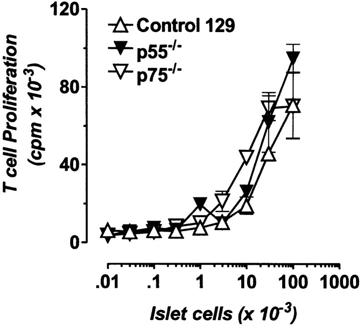
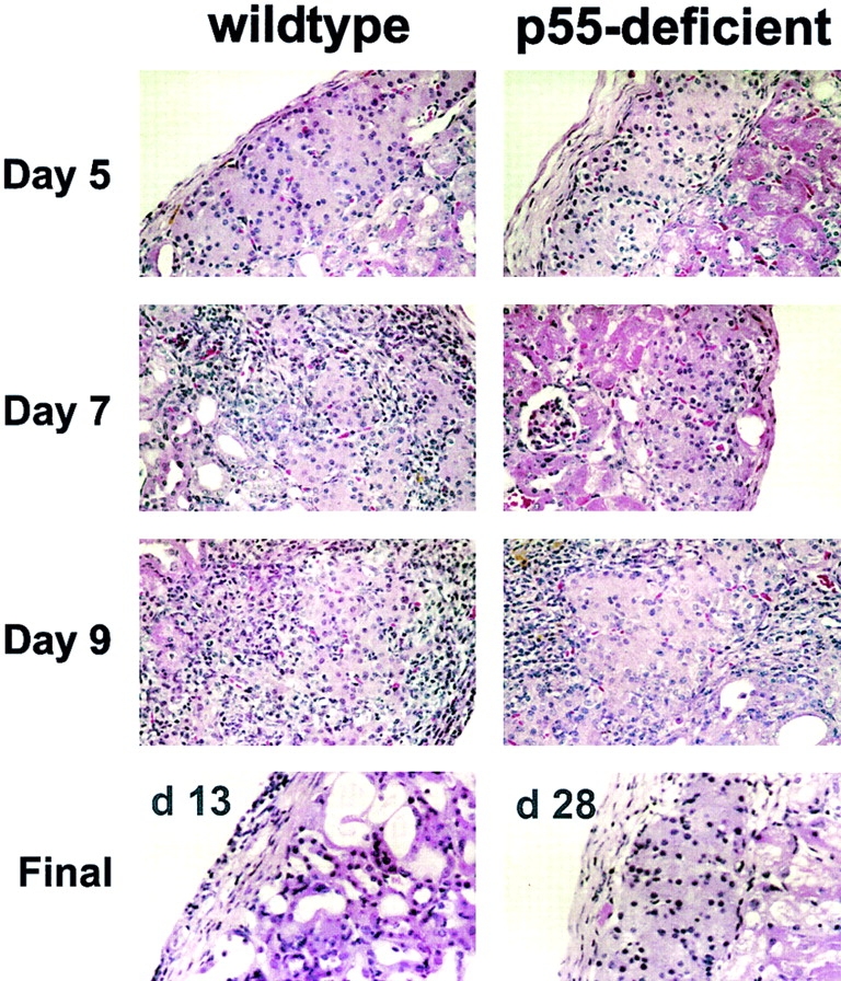
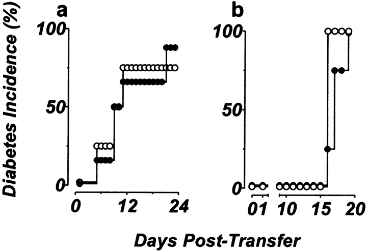
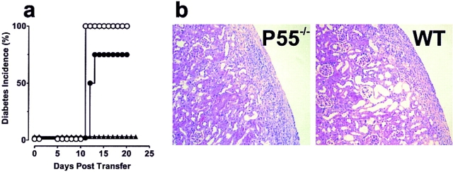
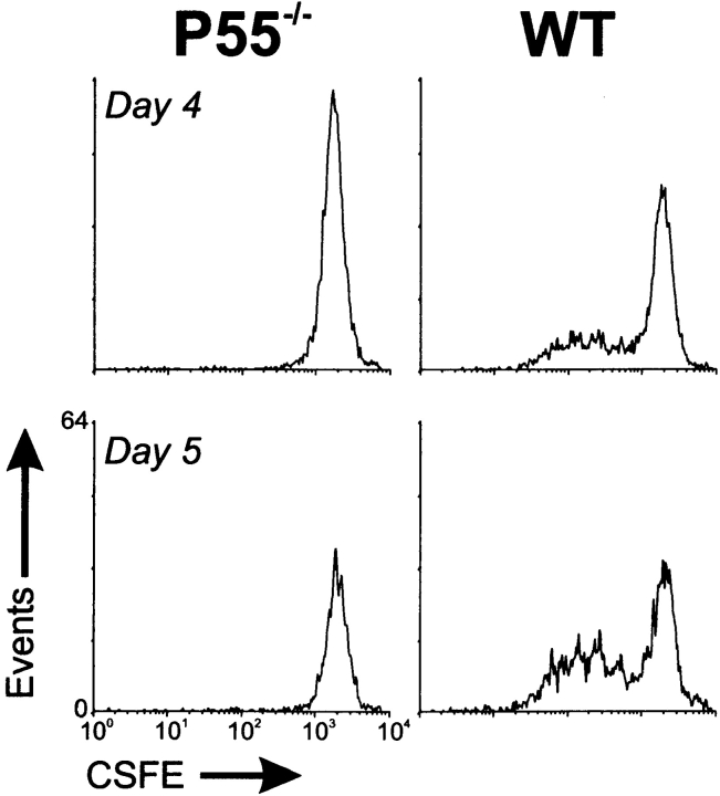
The islet-infiltrating and disease-causing leukocytes that are a hallmark of insulin-dependent diabetes mellitus produce and respond to a set of cytokine molecules. Of these, interleukin 1beta, tumor necrosis factor (TNF)-alpha, and interferon (IFN)-gamma are perhaps the most important. However, as pleiotropic molecules, they can impact the path leading to beta cell apoptosis and diabetes at multiple points. To understand how these cytokines influence both the formative and effector phases of insulitis, it is critical to determine their effects on the assorted cell types comprising the lesion: the effector T cells, antigen-presenting cells, vascular endothelium, and target islet tissue. Here, we report using nonobese diabetic chimeric mice harboring islets deficient in specific cytokine receptors or cytokine-induced effector molecules to assess how these compartmentalized loss-of-function mutations alter the events leading to diabetes. We found that islets deficient in Fas, IFN-gamma receptor, or inducible nitric oxide synthase had normal diabetes development; however, the specific lack of TNF- alpha receptor 1 (p55) afforded islets a profound protection from disease by altering the ability of islet-reactive, CD4(+) T cells to establish insulitis and subsequently destroy islet beta cells. These results argue that islet cells play a TNF-alpha-dependent role in their own demise.


Figures

Figure 1
Figure 1
Schematic representation of transplantation protocol. 6–8-wk-old healthy NOD.scid mice were injected with Streptozotocin to render them chemically diabetic (>400 mg/dl). Within 48–72 h of the onset of diabetes, the mice were engrafted with 250–300 islets under the left kidney capsule. Normoglycemia returned within 24 h of islet transplantation. The mice were then rested 7–10 d, during which time host-derived vascular support for the grafts developed. Splenic T cells from overtly diabetic BDC2.5/NOD.scid mice were transferred into the engrafted-NOD.scid mice (day 0). Mice were followed thereafter for the onset of diabetes. At day 28, normoglycemic mice were nephrectomized.
Figure 2
Figure 2
TNF-αRI (p55)–deficient islets are protected from destruction by diabetogenic CD4+ T cells, whereas Fas-deficient, IFN-γR–deficient, iNOS-deficient, and TNF-αR p75 islets are destroyed. (a) 250 B6.lpr/lpr (•, n = 12) or control B6 islets (○, n = 10) transplanted under the kidney capsule are destroyed. □, diabetes in control NOD.scid mice transferred with the same population of T cells (n = 9). (b) 250 IFN-γR– deficient (♦, n = 7), iNOS-deficient (▪, n = 6) or control 129 islets (▵, n = 8) transplanted under the kidney capsule are destroyed. (c) Transplanted p55−/− islets (▾, n = 9) and p55−/−p75−/− doubly deficient islets (▴, n = 10) are protected from destruction by transferred T cells from diabetic BDC2.5/NOD.scid mice, whereas p75−/− islets (▿, n = 11) are destroyed. Diabetes is measured by sampling venous blood using a standard one-step glucometer. Mice are considered diabetic after two successive readings ≥250 mg/dl. In all cases, engrafted mice that did not receive diabetogenic T cells remained normoglycemic throughout the experimental period (>180 d); moreover, the engrafted islets were responsible for normoglycemia, as the hemi-nephrectomy of engrafted kidneys returned mice to hyperglycemia.
Figure 3
Figure 3
TNFR-deficient islet cells are antigenic to BDC2.5 T cells. 2 × 10⁴ BDC2.5 T cells were cocultured with 2.5 × 10⁵ irradiated NOD splenic APCs and varying numbers of either dispersed islet cells from control 129 (▵), p55-deficient (▾) or p75-deficient (▿) donors. After 72 h, the assay was pulsed with [3H]TdR for 6 h to measure T cell proliferation to antigen. Data are depicted as mean ± SD.
Figure 4
Figure 4
TNFR (p55) expression on the islet mass is necessary for sustained infiltration of islets. Histological analysis of the progression of insulitis in p55-deficient islet grafts and control islet grafts (p75-deficient or 129-negative littermates) at the indicated time after transfer of T cells from diabetic BDC2.5/NOD.scid mice. Engrafted kidneys were fixed in buffered 10% formalin, embedded, and sectioned (4 μm). Sections were stained with hematoxylin and eosin to show infiltration and islet architecture. A representative image from one of three mice at each time point is shown. Original magnification: ×400.
Figure 5
Figure 5
Reciprocal islet transplants and mixed transplants of p55- deficient and p55-sufficient islets are destroyed upon BDC2.5 T cell infiltration. (a) Reciprocal transplants of p55-sufficient islets transplanted under the kidney capsule of p55−/− NOD.scid mice (•, n = 9) are destroyed as efficiently as p55+/+ islets transplanted under the kidney capsule of p55+/− NOD.scid mice (○, n = 4). (b) Both control islet grafts (○; 300 islets; n = 4) and mixed islet grafts (•; 200 p55-deficient islets and 100 control islets; n = 8) are destroyed with similar kinetics upon transfer of T cells from diabetic BDC2.5/NOD.scid mice. Diabetes is measured by sampling venous blood using a standard one-step glucometer. Mice are considered diabetic after two successive readings ≥250 mg/dl.
Figure 6
Figure 6
Spatial separation of the p55-deficient and p55-sufficient islet grafts on opposing kidneys does not protect the p55-deficient grafts from destruction. (a) In dual-kidney grafts, control NOD.scid mice were engrafted with p55-sufficient islet grafts under both kidneys (○; 200 islets on left kidney and 100 islets on the right) and experimental mice were engrafted with 200 p55-deficient islets on the left kidney and 100 p55-sufficient islets on the right kidney (•). Both sets of mice received T cells from BDC2.5/NOD.scid mice. In both cases the engrafted islets were destroyed with similar kinetics. Mice engrafted with p55-deficient islets only (▴) did not develop diabetes. Diabetes is measured by sampling venous blood using a standard one-step glucometer. Mice are considered diabetic after two successive readings ≥250 mg/dl. (b) Photomicrographs of the contralateral kidney grafts 14 d after transfer of T cells. Engrafted kidneys were fixed in buffered 10% formalin, embedded, and sectioned (4 μm). Sections were stained with hematoxylin and eosin to show infiltration and islet architecture. Note that the grafts are destroyed and only show residual scar tissue remains of both the p55-deficient graft, left, and the p55-sufficient graft, right. Representative images are from one of four mice analyzed at this time point. Original magnification: ×400.
Figure 7
Figure 7
T cells from the efferent lymph of p55−/− islet engrafted mice have not undergone cell division, whereas those from wild-type islet engrafted mice have. Streptozotocin-treated NOD.scid mice were engrafted with either p55-deficient (p55−/−) or p55-sufficient islets (WT) under the left kidney capsule. Several weeks later, 107 CSFE-labeled BDC2.5 T cells were injected intravenously in each group of mice. On days 4 and 5 after transfer, peri-renal lymph nodes were collected and BDC2.5 T cell proliferation was assessed as measured by diminution of CSFE label on a flow cytometer. Lymph nodes from two to four animals were pooled per group.

References

    1. Bach JF. Insulin-dependent diabetes mellitus as an autoimmune disease. Endocr Rev. 1994;15:516–542. - PubMed
    1. Atkinson MA, MacLaren NK. The pathogenesis of insulin-dependent diabetes mellitus. N Engl J Med. 1994;331:1428–1436. - PubMed
    1. Tisch R, McDevitt H. Insulin-dependent diabetes mellitus. Cell. 1996;85:291–297. - PubMed
    1. Charlton B, Bacelj A, Mandel TE. Administration of silica particles or anti-Lyt2 antibody prevents β-cell destruction in NOD mice given cyclophosphamide. Diabetes. 1988;37:930–935. - PubMed
    1. Miller BJ, Appel MC, O'Neil J, Wicker LS. Both the LYT-2+ and L3T4+T cell subsets are required for transfer of diabetes in nonobese diabetic mice. J Immunol. 1988;140:52–58. - PubMed

Publication types

MeSH terms