. 1999 Apr 5;189(7):1073-81.
doi: 10.1084/jem.189.7.1073.

Bone marrow NK1.1(-) and NK1.1(+) T cells reciprocally regulate acute graft versus host disease

Affiliations

Bone marrow NK1.1(-) and NK1.1(+) T cells reciprocally regulate acute graft versus host disease

D Zeng et al. J Exp Med. .

Abstract

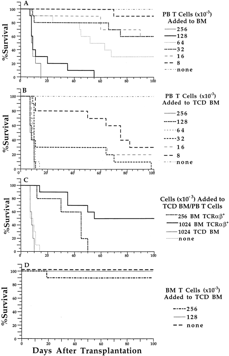
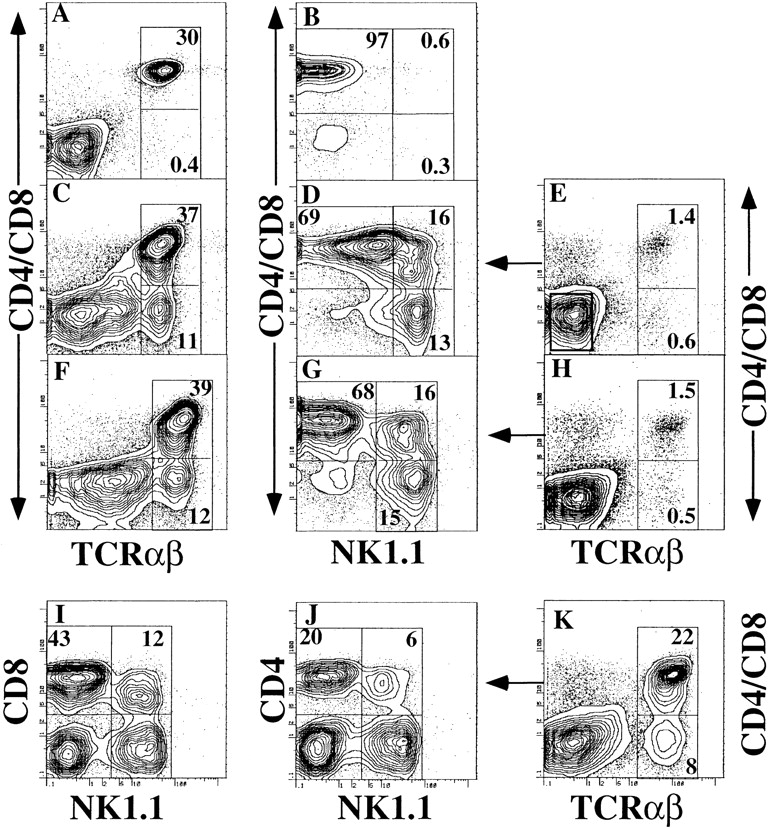
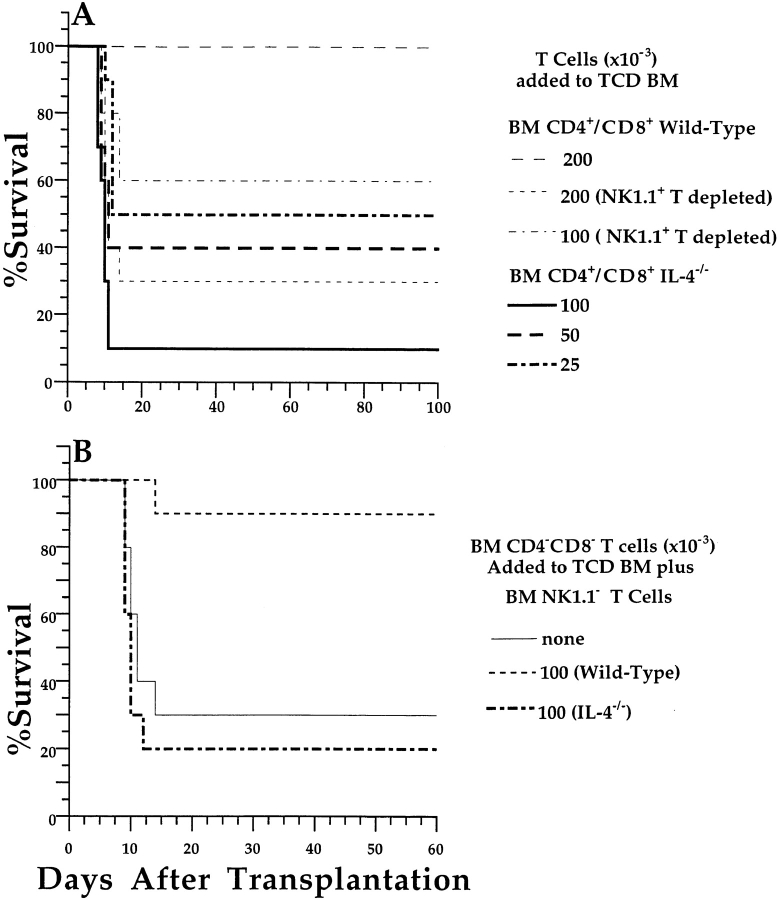
Sorted CD4(+) and CD8(+) T cells from the peripheral blood or bone marrow of donor C57BL/6 (H-2(b)) mice were tested for their capacity to induce graft-versus-host disease (GVHD) by injecting the cells, along with stringently T cell-depleted donor marrow cells, into lethally irradiated BALB/c (H-2(d)) host mice. The peripheral blood T cells were at least 30 times more potent than the marrow T cells in inducing lethal GVHD. As NK1.1(+) T cells represented <1% of all T cells in the blood and approximately 30% of T cells in the marrow, the capacity of sorted marrow NK1.1(-) CD4(+) and CD8(+) T cells to induce GVHD was tested. The latter cells had markedly increased potency, and adding back marrow NK1.1(+) T cells suppressed GVHD. The marrow NK1.1(+) T cells secreted high levels of both interferon gamma (IFN-gamma) and interleukin 4 (IL-4), and the NK1.1(-) T cells secreted high levels of IFN-gamma with little IL-4. Marrow NK1.1(+) T cells obtained from IL-4(-/-) rather than wild-type C57BL/6 donors not only failed to prevent GVHD but actually increased its severity. Together, these results demonstrate that GVHD is reciprocally regulated by the NK1.1(-) and NK1.1(+) T cell subsets via their differential production of cytokines.


Figures

Figure 1
Figure 1
Ability of peripheral blood (PB) and bone marrow (BM) CD4+ and CD8+ T cells to induce lethal GVHD. Graded numbers of sorted CD4+/CD8+ T cells were added to a constant number (1.5 × 10⁶) of unfractionated or TCD C57BL/6 marrow cells and injected intravenously into lethally irradiated (800 cGy) BALB/c hosts. Survival over a 100-d observation period is shown in groups of 10 mice. (A) Graded numbers of sorted peripheral blood CD4+/CD8+ T cells were added to unfractionated marrow cells. (B) Graded number of sorted peripheral blood CD4+/CD8+ T cells were added to TCD marrow cells. (C) 6.4 × 10⁴ peripheral blood CD4+/CD8+ T cells and 1.5 × 10⁶ TCD marrow cells were or were not injected with sorted TCRα/β+ marrow T cells or with TCD marrow cells. (D) Sorted CD4+/CD8+ T cells from the marrow were injected with 1.5 × 10⁶ TCD marrow cells.
Figure 2
Figure 2
Two-color flow cytometric analysis of α/β+ T cells in the peripheral blood, bone marrow, and T cell–enriched bone marrow of C57BL/6 wild-type or C57BL IL-4−/− mice. A and B show staining of wild-type PBMC. (A) CD4 and CD8 (using same fluorochrome) versus TCRαβ markers; the two boxes enclose CD4+/CD8+ (upper) and CD4CD8 (lower) TCRαβhi cells. (B) Analysis of the gated TCRαβhi cells for CD4 and CD8 versus NK1.1. Left upper box encloses NK1.1 CD4+/CD8+ cells, right upper box encloses NK1.1+ CD4+/CD8+ cells, and right lower box encloses NK1.1+ CD4CD8 cells. C and D show similar analyses for immunomagnetic bead–enriched wild-type bone marrow T cells. F and G show analyses of enriched IL-4−/− bone marrow T cells. E shows analysis of whole wild-type bone marrow cells before enrichment of T cells and thresholds for gating TCD bone marrow cells (E, left box). Sorted TCRαβ+ bone marrow cells were all cells to the right of the TCRαβ gating threshold. H shows analysis of whole bone marrow cells before enrichment from IL-4−/− mice. K shows analysis of enriched bone marrow cells from wild-type mice; box encloses TCRαβhi cells. I and J show the analyses of the gated TCRαβhi cells for CD8 or CD4 versus NK1.1, respectively. Percentages of cell subsets enclosed in boxes are shown.
Figure 3
Figure 3
Different subsets of T cells induce or suppress GVHD, depending on their cytokine secretion profiles. (A) Sorted C57BL/6 wild-type CD4+ and CD8+ bone marrow (BM) T cells with or without depletion of NK1.1+ T cells were added to C57BL/6 wild-type TCD bone marrow cells (1.5 × 10⁶) and injected into irradiated BALB/c hosts. In addition, sorted C57BL IL-4−/− CD4+ and CD8+ bone marrow T cells without NK1.1+ T cell depletion were added to wild-type TCD bone marrow cells and injected into BALB/c hosts. (B) Sorted CD4CD8 T cells from the bone marrow of wild-type or IL-4−/− C57BL/6 donor mice were added to 2.00 × 10⁵ sorted wild-type NK1.1 CD4+/CD8+ bone marrow T cells and 1.5 × 10⁶ wild-type TCD bone marrow cells. Control mice received sorted wild-type NK1.1 CD4+/CD8+ bone marrow T cells and TCD bone marrow cells without added-back CD4CD8+ bone marrow T cells (none). There were 10 mice in each group.
Figure 4
Figure 4
Different subsets of NK1.1 and NK1.1+ T cells induce or suppress histopathological lesions of GVHD in the large intestine and skin. Sections of intestine (A) and skin (B) from irradiated BALB/c hosts injected 60 d earlier with only TCD bone marrow from wild-type C57BL/6 donor mice. Plump, mucin-containing epithelial cells are seen in the intestine (arrows) with little or no inflammation. C and D show BALB/c hosts injected with wild-type TCD bone marrow and sorted wild-type NK1.1 CD4+/ CD8+ bone marrow T cells. An inflammatory infiltrate between intestinal crypts and hyperplasia of crypt epithelial cells with reduced mucin characteristic of GVHD is seen (arrow). An inflammatory infiltrate in the dermis and epidermal hyperplasia characteristic of GVHD is observed in the skin (arrows). E and F show hosts injected with wild-type TCD bone marrow, sorted wild-type NK1.1 CD4+/CD8+ bone marrow T cells, and sorted wild-type CD4CD8 (NK1.1+) bone marrow T cells. Lesions of GVHD are markedly reduced. G and H show hosts injected with wild-type TCD bone marrow, sorted wild-type NK1.1 CD4+/CD8+ bone marrow T cells, and sorted IL-4−/− CD4CD8 (NK1.1+) bone marrow T cells. Severe lesions of GVHD are seen, including an abscess in an intestinal crypt (arrow).

References

    1. Sullivan, K.M. 1994. Graft-versus-host disease. In Bone Marrow Transplantation. S.J. Forman, K.G. Blume, and E.D. Thomas, editors. Blackwell Scientific Publications, Boston. xxiii, 942.
    1. Weisdorf D, Hakke R, Blazar B, Miller W, McGlave P, Ramsay N, Kersey J, Filipovich A. Risk factors for acute graft-versus-host disease in histocompatible donor bone marrow transplantation. Transplantation. 1991;51:1197–1203. - PubMed
    1. Bross DS, Tutschka PJ, Farmer ER, Beschorner WE, Braine HG, Mellits ED, Bias WB, Santos GW. Predictive factors for acute graft-versus-host disease in patients transplanted with HLA-identical bone marrow. Blood. 1984;63:1265–1270. - PubMed
    1. Ferrara JL, Cooke KR, Pan L, Krenger W. The immunopathophysiology of acute graft-versus-host-disease. Stem Cells. 1996;14:473–489. - PubMed
    1. Korngold R, Sprent J. Surface markers of T cells causing lethal graft-vs-host disease to class I vs class II H-2 differences. J Immunol. 1985;135:3004–3010. - PubMed

Publication types

MeSH terms