

Mandarin and English single word processing studied with functional magnetic resonance imaging
- PMID: 10191322
- PMCID: PMC6782281
- DOI: 10.1523/JNEUROSCI.19-08-03050.1999
Mandarin and English single word processing studied with functional magnetic resonance imaging
Abstract
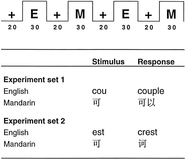
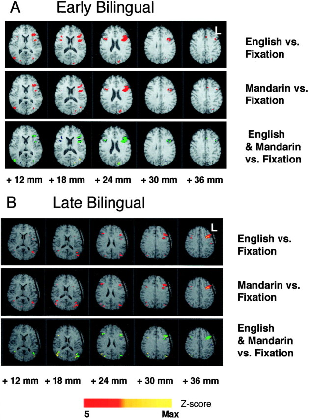
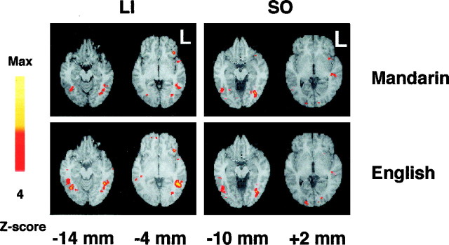
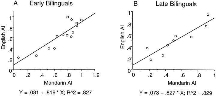
The cortical organization of language in bilinguals remains disputed. We studied 24 right-handed fluent bilinguals: 15 exposed to both Mandarin and English before the age of 6 years; and nine exposed to Mandarin in early childhood but English only after the age of 12 years. Blood oxygen level-dependent contrast functional magnetic resonance imaging was performed while subjects performed cued word generation in each language. Fixation was the control task. In both languages, activations were present in the prefrontal, temporal, and parietal regions, and the supplementary motor area. Activations in the prefrontal region were compared by (1) locating peak activations and (2) counting the number of voxels that exceeded a statistical threshold. Although there were differences in the magnitude of activation between the pair of languages, no subject showed significant differences in peak-location or hemispheric asymmetry of activations in the prefrontal language areas. Early and late bilinguals showed a similar pattern of overlapping activations. There are no significant differences in the cortical areas activated for both Mandarin and English at the single word level, irrespective of age of acquisition of either language.
Figures
References
-
- Albert M, Obler L. The bilingual brain: neuropsychological and neurolinguistic aspects of bilingualism. Academic; New York: 1978.
-
- Binder JR, Swanson SJ, Hammeke TA, Morris GL, Mueller WM, Fischer M, Benbadis S, Frost JA, Rao SM, Haughton VM. Determination of language dominance using functional MRI: a comparison with the WADA test. Neurology. 1996;46:978–984. - PubMed
-
- Buchel C, Price C, Friston K. A multimodal language region in the ventral visual pathway. Nature. 1998;394:274–277. - PubMed
-
- Buckner RL, Raichle ME, Petersen SE. Dissociation of human prefrontal cortical areas across different speech production tasks and gender groups. J Neurophysiol. 1995;74:2163–2173. - PubMed
Publication types
MeSH terms
Substances
LinkOut - more resources
Full Text Sources
Medical