. 1999 Apr 15;19(8):3171-82.
doi: 10.1523/JNEUROSCI.19-08-03171.1999.

Hypothalamic hypocretin (orexin): robust innervation of the spinal cord

Affiliations

Hypothalamic hypocretin (orexin): robust innervation of the spinal cord

A N van den Pol. J Neurosci. .

Abstract

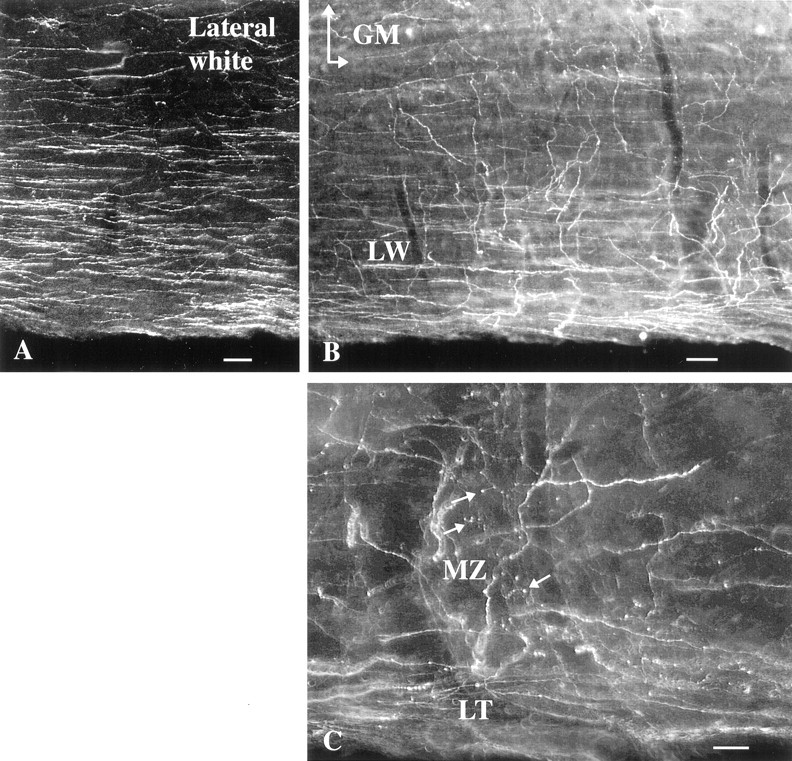
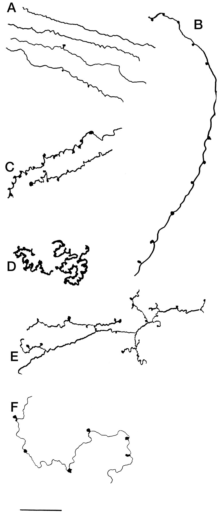
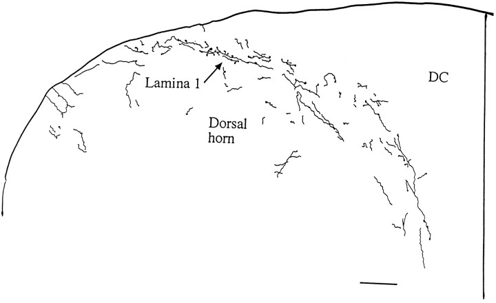
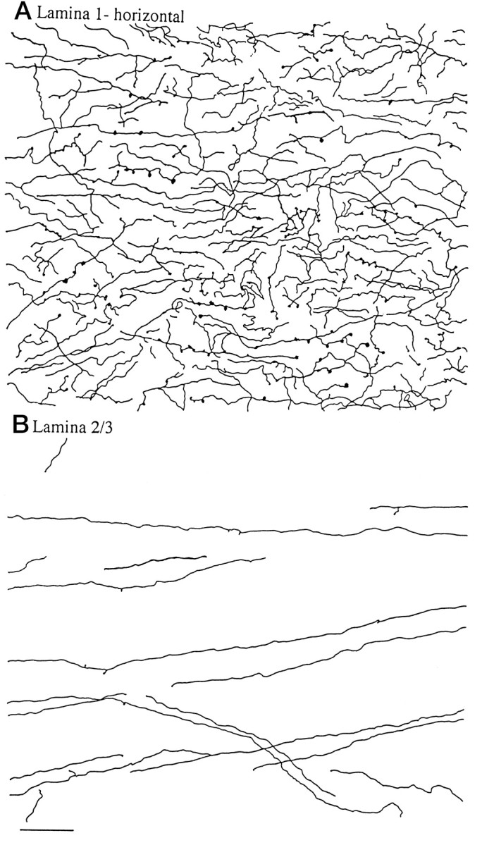
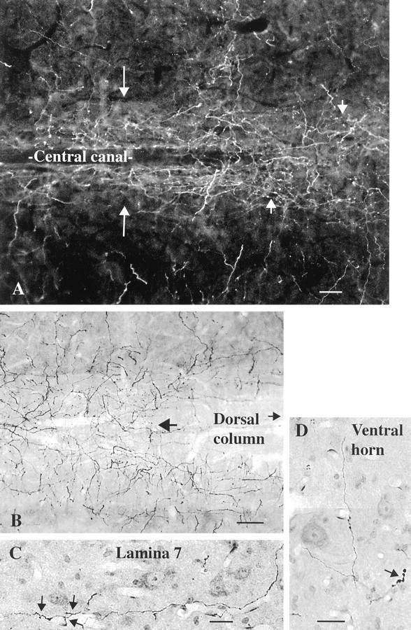
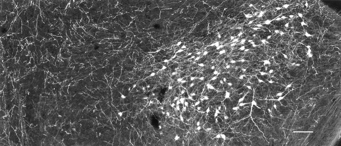
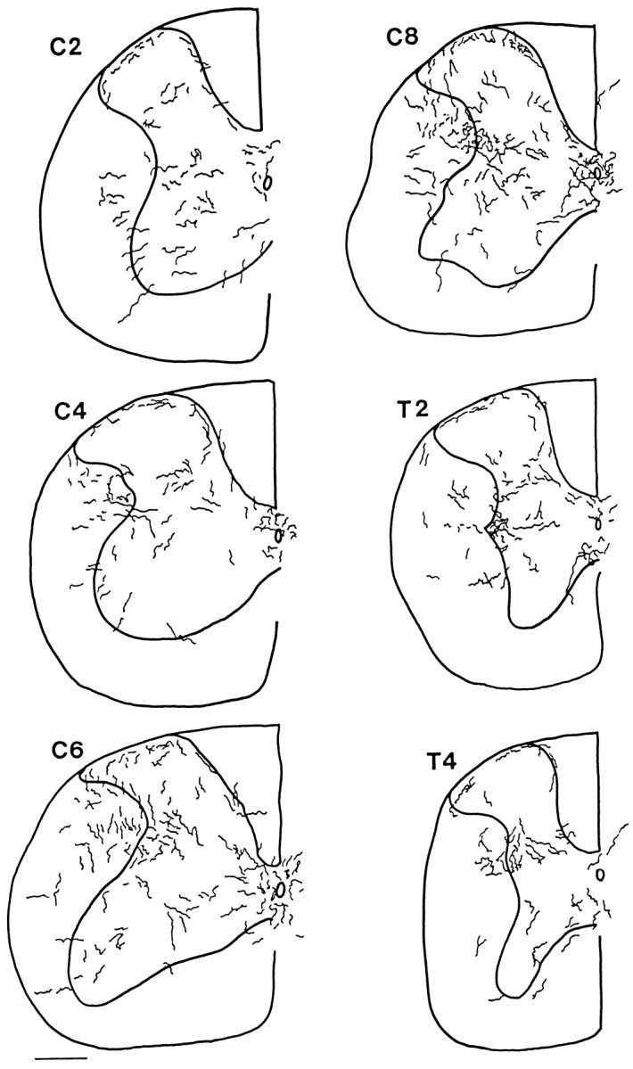
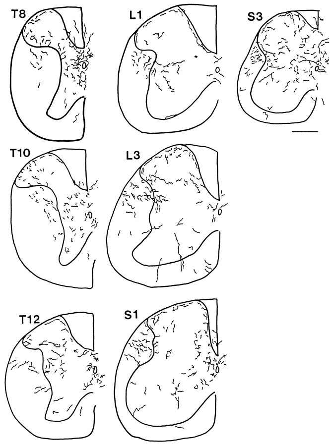
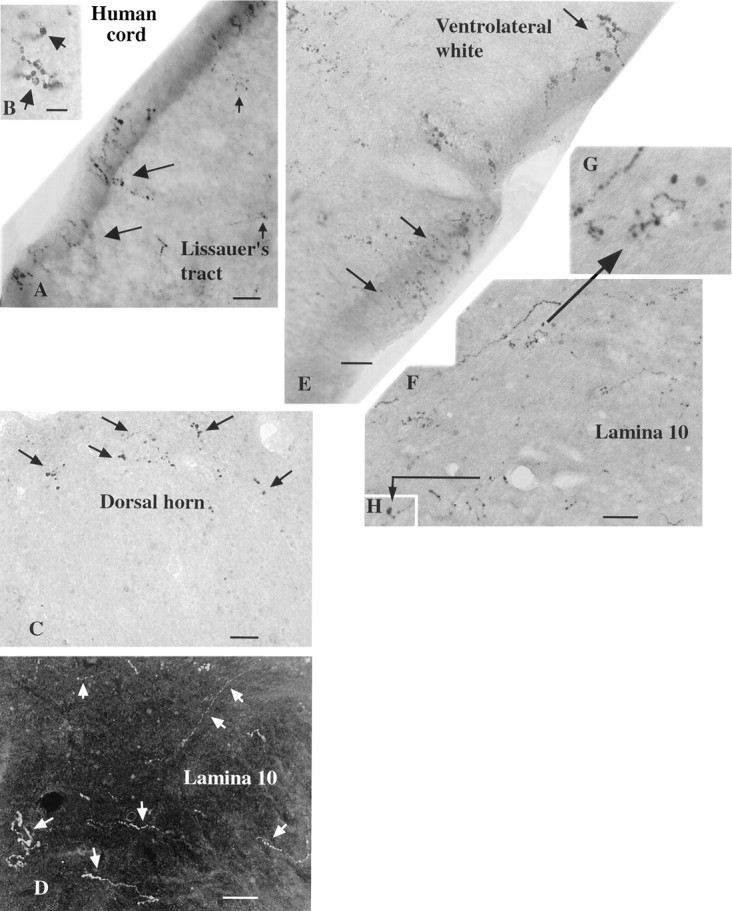
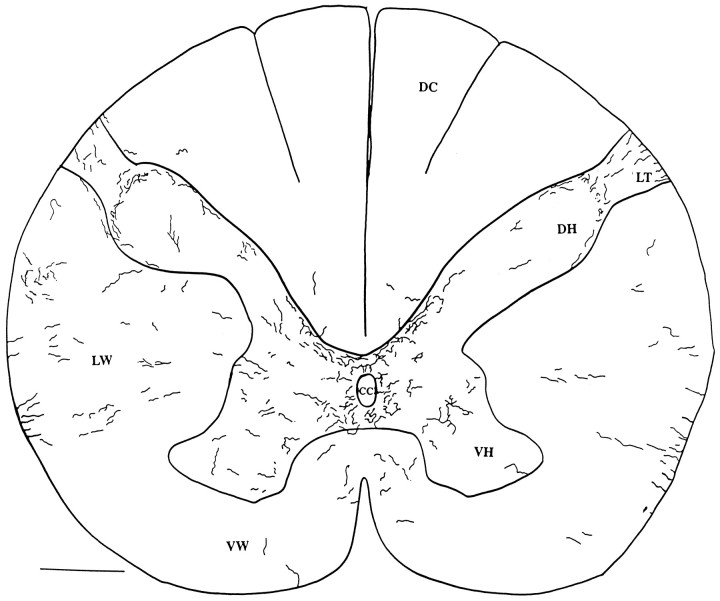
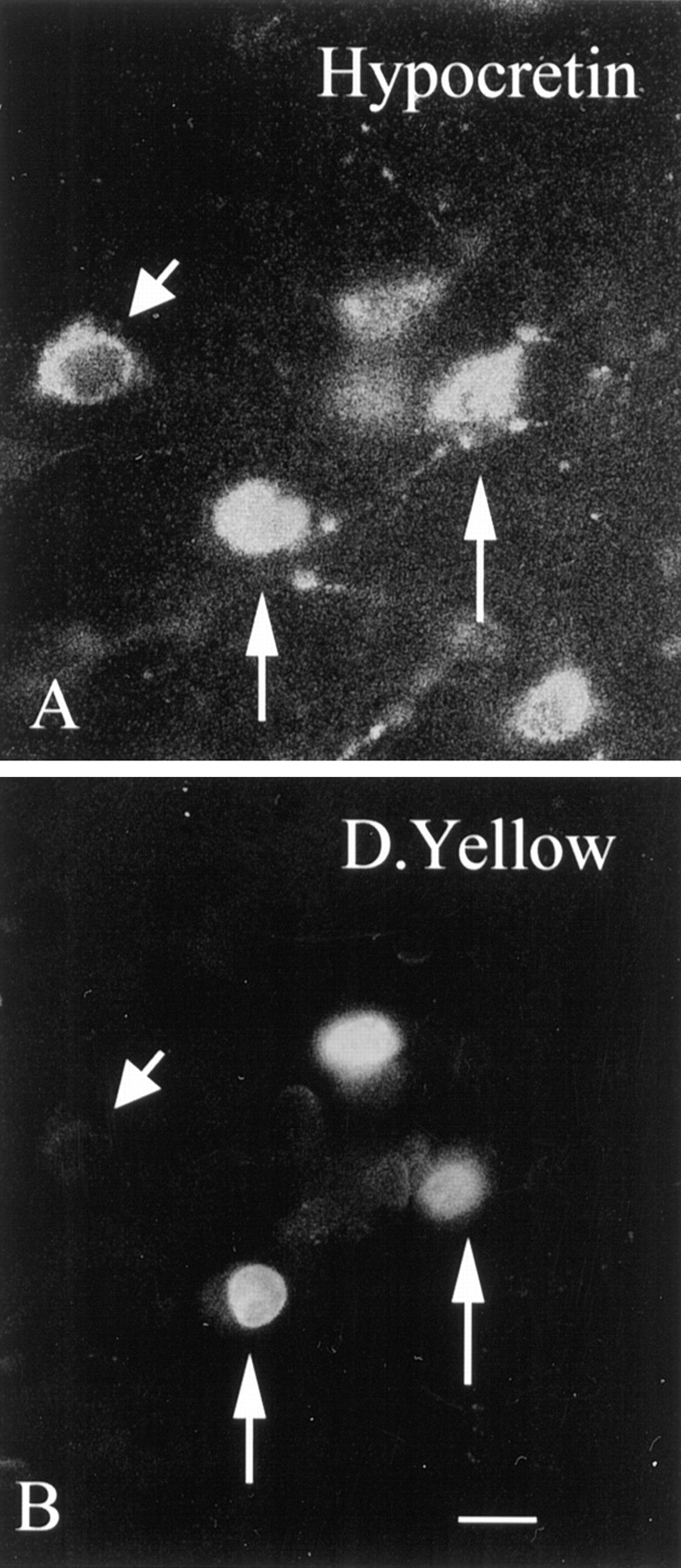
Hypocretin (orexin) is synthesized by neurons in the lateral hypothalamus and has been reported to increase food intake and regulate the neuroendocrine system. In the present paper, long descending axonal projections that contain hypocretin were found that innervate all levels of the spinal cord from cervical to sacral segments, as studied in mouse, rat, and human spinal cord and not previously described. High densities of axonal innervation are found in regions of the spinal cord related to modulation of sensation and pain, notably in the marginal zone (lamina 1). Innervation of the intermediolateral column and lamina 10 as well as strong innervation of the caudal region of the sacral cord suggest that hypocretin may participate in the regulation of both the sympathetic and parasympathetic parts of the autonomic nervous system. Double-labeling experiments in mice combining retrograde transport of diamidino yellow after spinal cord injections and immunocytochemistry support the concept that hypocretin-immunoreactive fibers in the cord originate from the neurons in the lateral hypothalamus. Digital-imaging physiological studies with fura-2 detected a rise in intracellular calcium in response to hypocretin in cultured rat spinal cord neurons, indicating that spinal cord neurons express hypocretin-responsive receptors. A greater number of cervical cord neurons responded to hypocretin than another hypothalamo-spinal neuropeptide, oxytocin. These data suggest that in addition to possible roles in feeding and endocrine regulation, the descending hypocretin fiber system may play a role in modulation of sensory input, particularly in regions of the cord related to pain perception and autonomic tone.


Figures

Fig. 1.
Fig. 1.
Hypocretin-immunoreactive axons in rat white matter, horizontal section. A, In the dorsal region of the lateral white matter, a high density of immunoreactive axons is found in this dark-field micrograph. B, More ventrally in the lateral white matter (LW), some axons turn medially to enter the gray matter (GM,top). C, In the dorsal part of the cord in the region of Lissauer’s tract (LT), some axons leave a group of descending axons at the bottomand enter into part of the marginal zone (MZ) where boutons are found (arrows). Scale bars:A, 30 μm; B, 25 μm; C, 15 μm.
Fig. 2.
Fig. 2.
Hypocretin axons with a variety of shapes in rat cord. A, Thin parallel axons entering the gray matter from the dorsolateral white matter. B, Long axons descend from lamina 3/4 down to lamina 7 with boutons at regular intervals in each lamina the axon traverses. C, Axons may traverse the intermediolateral column with frequent boutons.D, Some axons are thick with large numbers of tightly packed boutons investing a small region of lamina 5. E, Lateral to the central canal, axons have frequent branches.F, In the ventral horn, thin axons maintain only a few boutons. Scale bar, 20 μm.
Fig. 3.
Fig. 3.
Lamina 1 of the dorsal horn in rat. A single 50-μm-thick section was used to trace hypocretin-immunoreactive axons in a cross-section of the midcervical dorsal horn. Boutons were particularly prevalent in lamina 1. DC, Dorsal columns. Scale bar, 70 μm.
Fig. 4.
Fig. 4.
Dorsal horn, horizontal sections in rat.A, A substantial number of hypocretin-immunoreactive axons are found in 40-μm-thick horizontal sections of the midcervical cord, shown here from camera lucida drawings. B, In contrast to the marginal zone above, in laminae 2 and 3, relatively few axons are found, and those that do appear show little branching and few boutons. Both regions were drawn with the same size dimensions. Scale bar, 45 μm.
Fig. 5.
Fig. 5.
Hypocretin-immunoreactive axons in rat gray matter, horizontal section. A, In the midcord, the highest density of hypocretin-immunoreactive axons is found around the central canal (long arrows). The short arrows indicate axons in lamina 10 immediately above the central canal. B, At a slightly higher plane of horizontal section, the ventral aspect of the dorsal columns is seen and continues to the right in the direction of thesmall arrow. On the right, just under the dorsal column white matter (large arrow), the axon density increases. C, In lamina 7, a hypocretin-immunoreactive axon and some of its collaterals (arrows) are found among cells stained with nuclear red.D, In the ventral horn, occasional axons are found, sometimes with swellings, as indicated by the arrow. Scale bars: A, 20 μm; B, 22 μm;C, D, 18 μm.
Fig. 6.
Fig. 6.
Hypocretin-immunoreactive perikarya in mouse. In the lateral hypothalamus and perifornical area, hypocretin-immunoreactive cell bodies and their processes are labeled. The midline is on the left, and lateral is on theright. Scale bar, 60 μm.
Fig. 7.
Fig. 7.
Hypocretin axons in mouse spinal cord, C2–T4. Camera lucida drawings were made in regular intervals from 30 μm cross-sections. The outline of the cord and the outline of the gray matter are indicated by the thick lines. To allow visibility of axons when reduced in size, the diameter of the axons is greater on the drawing than it actually appeared in the microscope. Axons that were descending the cord, particularly in dorsolateral white matter, and oriented parallel with the long axis of the cord, which consequently would appear only as a single point in these cross-sections, were not included.
Fig. 8.
Fig. 8.
Hypocretin axons in mouse spinal cord, T6–S4. Continuation of the previous figure.
Fig. 9.
Fig. 9.
Hypocretin-immunoreactive axons in human spinal cord, photomicrographs. A, In the dorsal region of the human cervical cord, immunoreactive axons (large andsmall arrows) are found at the surface of the cord and down into Lissauer’s tract. B, Higher magnification of an axon in the same region as A. C, In the dorsal horn the immunoreactive axons (arrows) form a semicircle in the area of lamina 1. D, In lamina 10 a number of thin and thicker axons are found (arrows) in this dark-field micrograph. E, Immunoreactive axons (arrows) travel to the edge of the cord in the ventrolateral white matter. The beaded appearance of immunoreactive axons in the human cord is probably attributable to inadequate immersion fixation, which may result in some axonal degeneration before axonal stabilization. F, In lamina 10 axons and boutons are found. Two sets of small terminal boutons are shown at higher magnification in G and H, as indicated by the arrows. Scale bars: A, E, 12 μm;B, 7 μm; C, 20 μm; D, 18 μm; H, 15 μm.
Fig. 10.
Fig. 10.
Human cervical spinal cord. Camera lucida drawing was made of hypocretin-immunoreactive axons in two adjacent 40 μm sections of a C4–C5 cross-section of a human cord. DH, Dorsal horn; VH, ventral horn; DC, dorsal columns; cc, central canal; LW, lateral white matter; VW, ventral white matter;LT, Lissauer’s tract. Scale bar, 1 mm.
Fig. 11.
Fig. 11.
Retrograde transport of tracer to hypocretin-immunoreactive cells in lateral hypothalamus. In the same section, neurons immunoreactive for hypocretin (A) and also labeled with diamidino yellow (D.Yellow) (B) transported from the spinal cord back to the hypothalamus. The two vertical arrowsin A and B show hypocretin-immunoreactive neurons in which the nucleus is labeled with diamidino yellow. Theshort arrowhead shows a hypocretin-immunoreactive neuron in the same field that shows no nuclear labeling for diamidino yellow. Scale bar, 5 μm.
Fig. 12.
Fig. 12.
Spinal cord neurons show calcium rise in response to hypocretin. A, Hypocretin-2 (H, 1 μm) was added by bath perfusion. In the presence of hypocretin, cytoplasmic calcium increased as measured with fura-2 imaging. Horizontal lines mark the presence of the peptide. B, A neuron responds both to hypocretin-1 (H-1) and hypocretin-2 (H-2) but not to the 17-amino acid inactive C-terminal fragment (17) of preprohypocretin. C, D, In two neurons recorded simultaneously, the response to hypocretin-2 (H, 1 μm) and oxytocin (Oxy, 1 μm) was compared. InC, the neuron responds to hypocretin but shows little response to oxytocin. The neuron in D responds to both oxytocin and hypocretin.

Similar articles

Cited by

References

    1. Armstrong WE, Warach S, Hatton GI, NcNeill TH. Subnuclei in the rat paraventricular nucleus: a cytoarcitectural, HRP, and immunocytochemical analysis. Neuroscience. 1980;206:317–345. - PubMed
    1. Bittencourt JC, Elias CF. Melanin-concentrating hormone and neuropeptide EI projections from the lateral hypothalamic area and zona incerta to the medial septal nucleus and spinal cord: a study using multiple neuronal tracers. Brain Res. 1998;805:1–19. - PubMed
    1. Bittencourt JC, Presse F, Arias C, Vaughan J, Nahon JL, Vale W, Sawchenko PE. The melanin-concentrating hormone system of the rat brain: an immuno- and hybridization histochemical characterization. J Comp Neurol. 1992;319:218–245. - PubMed
    1. Brown AG. Organization in the spinal cord. The anatomy and physiology of identified neurons, pp 1–238. Springer; New York: 1981.
    1. Cedarbaum JM, Aghajanian GK. Afferent projections to the rat locus coeruleus as determined by a retrograde tracing technique. J Comp Neurol. 1978;178:1–16. - PubMed

LinkOut - more resources