. 1999 Feb;126(4):849-58.
doi: 10.1038/sj.bjp.0702348.

Effects of the prostanoid EP3-receptor agonists M&B 28767 and GR 63799X on infarct size caused by regional myocardial ischaemia in the anaesthetized rat

Affiliations

Effects of the prostanoid EP3-receptor agonists M&B 28767 and GR 63799X on infarct size caused by regional myocardial ischaemia in the anaesthetized rat

K Zacharowski et al. Br J Pharmacol. 1999 Feb.

Abstract

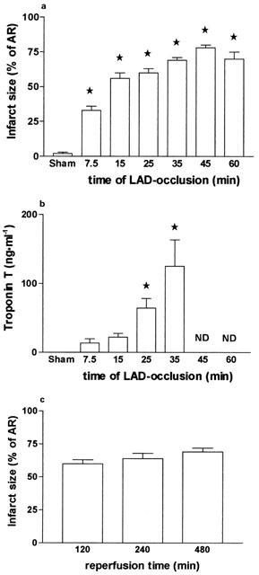
1. This study investigates the effects of two agonists of the prostanoid EP3-receptor (M&B 28767 and GR 63799X) on the infarct size caused by regional myocardial ischaemia and reperfusion in the anaesthetized rat. 2. One hundred and sixty-seven, male Wistar rats were anaesthetized (thiopentone, 120 mg kg(-1) i.p.), ventilated (8-10 ml kg(-1), 70 strokes min(-1), inspiratory oxygen concentration: 30%; PEEP: 1-2 mmHg) and subjected to occlusion of the left anterior descending coronary artery (LAD, for 7.5, 15, 25, 35, 45 or 60 min) followed by reperfusion (2 h). Infarct size was determined by staining of viable myocardium with a tetrazolium stain (NBT), histological evaluation by light and electron microscopy and determination of the plasma levels of cardiac troponin T. 3. M&B 28767 (0.5 microg kg(-1) min(-1), i.v., n=7) or GR 63799X (3 microg kg(-1) min(-1), i.v., n=7) caused significant reductions in infarct size from 60+/-3% (25 min ischaemia and 2 h reperfusion; saline-control, n=8) to 39+/-6 and 38+/-4% of the area at risk, without causing a significant fall in blood pressure. Pretreatment of rats with 5-hydroxydecanoate (5-HD), an inhibitor of ATP-sensitive potassium channels, attenuated the cardioprotective effects of both EP3-receptor agonists. The reduction in infarct size afforded by M&B 28767 was also abolished by glibenclamide and the protein kinase C (PKC) inhibitors staurosporine and chelerythrine. 4. Thus, M&B 28767 and GR 63799X reduce myocardial infarct size in the rat by a mechanism(s) which involves the activation of PKC and the opening of ATP-sensitive potassium channels.


Figures

Figure 1
Figure 1
(a) Infarct size (expressed as per cent of the area at risk, AR) caused by occlusion 7.5, 15, 25, 35, 45 or 60 min and followed by reperfusion (2 h) of the left anterior descending coronary artery (LAD) in the anaesthetized rat. (b) Alterations in the plasma levels of cardiac troponin T caused by occlusion for 7.5 15, 25 or 35 min and followed by reperfusion (2 h) of the left anterior descending coronary artery (LAD) in the anaesthetized rat. *P<0.05 when compared to sham-operated control. ND, not determined. (C) Infarct size (expressed as per cent of the area at risk, AR) caused by occlusion for 25 min and reperfusion for 2, 4 or 8 h of the left anterior descending coronary artery (LAD) in the anaesthetized rat.
Figure 2
Figure 2
(a) Histological section after Fuchsin staining of the area at risk of a rat heart subjected to 25 min of myocardial ischaemia followed by 2 h of reperfusion. Depicted is an example of complete coagulation necrosis with deeply eosinophilic cytoplasm of myocytes, which demonstrates a strong staining with Fuchsin. (b) Histological section after Luxol-Fast-Blue staining of the area at risk of a rat heart subjected to 25 min of myocardial ischaemia followed by 2 h of reperfusion. Depicted is a further example of complete coagulation necrosis with eosinophilic cytoplasm of myocytes which demonstrates a submaximal staining with Luxol-Fast-Blue in the peripheral myocytes.
Figure 3
Figure 3
Electron microscopical section of the area at risk of a rat heart subjected to 25 min of myocardial ischaemia followed by 2 h of reperfusion. (a) Depicted are typical examples of granular disruption of the I-bands and the Z-lines compared to a normal A-band. (b) Depicted is an example of early contraction bands, osmiophilic degradation of mitochondria and intramitochondrial calcifications. (c) Depicted are typical examples of swollen or osmophilic degraded mitochondria as well as vacuolization of the wall of small vessels. These findings are similar to those exhibited by sections of human heart at 12 h after transmural myocardial infarction.
Figure 3
Figure 3
Electron microscopical section of the area at risk of a rat heart subjected to 25 min of myocardial ischaemia followed by 2 h of reperfusion. (a) Depicted are typical examples of granular disruption of the I-bands and the Z-lines compared to a normal A-band. (b) Depicted is an example of early contraction bands, osmiophilic degradation of mitochondria and intramitochondrial calcifications. (c) Depicted are typical examples of swollen or osmophilic degraded mitochondria as well as vacuolization of the wall of small vessels. These findings are similar to those exhibited by sections of human heart at 12 h after transmural myocardial infarction.
Figure 4
Figure 4
Infarct size caused by occlusion (25 min) and reperfusion (2 h) of the left anterior descending coronary artery (LAD) in the anaesthetized rat. Different groups of animals were treated with (a) vehicle (control, n=8), M&B 28767 (0.5 μg kg−1 h−1, i.v., n=7), 5-hydroxydecanoate (5-HD, 5 mg kg−1, i.v., n=6), 5-HD plus M&B 28767 (n=6) and (b) vehicle (control, n=8), GR 63799X (3.0 μg kg−1, h−1, i.v., n=7), 5-HD (5 mg kg−1, i.v., n=6), 5-HD plus GR 63799X (n=6). *P<0.05 when compared to control.
Figure 5
Figure 5
Infarct size caused by occlusion (25 min) and reperfusion (2 h) of the left anterior descending coronary artery (LAD) in the anaesthetized rat. Different groups of animals were treated with (a) vehicle (control, n=8), M&B 28767 (0.5 μg kg−1 h−1, i.v., n=7), staurosporine (1 μg kg−1, i.v., n=6), staurosporine plus M&B 28767 (n=6) and (b) vehicle (control, n=8), M&B 28767 (0.5 μg kg−1 h−1, i.v., n=7), chelerythrine (0.7 mg kg−1, i.v., n=6), chelerythrine plus M&B 28767 (n=6) *P<0.05 when compared to control.

Similar articles

Cited by

References

    1. ADAMS J.E., ABENDSCHEIN D.R., JAFFE A.S. Biochemical markers of myocardial injury: is MB creatine kinase the choice for 1990s. Circ. 1993;88:750–763. - PubMed
    1. BALLER D., BRETSCHNEIDER H.J., HELLIGE G. A critical look at currently used indices of myocardial oxygen consumption. Basic Res. Cardiol. 1981;76:163–181. - PubMed
    1. BANCROFT J.D., STEVENS A. Theory and Practice of Histological Techniques 1995Edinburgh: Churchill Livingstone; ed. Bancroft, J.D. & Stevens, A
    1. BAROLDI G. Different types of myocardial necrosis in coronary heart disease: A pathophysiological review of their functional significance. Am. Heart. J. 1975;89:742–752. - PubMed
    1. COLEMAN R.A., KENNEDY I., HUMPHREY P.P.A., BUNCE K., LUMLEY P. Comprehensive Medicinal Chemistry 1990Oxford: Pergamon Press; 643ed. Hansch, C., Samnes, P.G., Taylor, J.B. & Emmett, J.C. p

Publication types