Review
. 1999 Apr;12(2):243-85.
doi: 10.1128/CMR.12.2.243.

Molecular techniques for detection, species differentiation, and phylogenetic analysis of microsporidia

Affiliations
Review

Molecular techniques for detection, species differentiation, and phylogenetic analysis of microsporidia

C Franzen et al. Clin Microbiol Rev. 1999 Apr.

Abstract

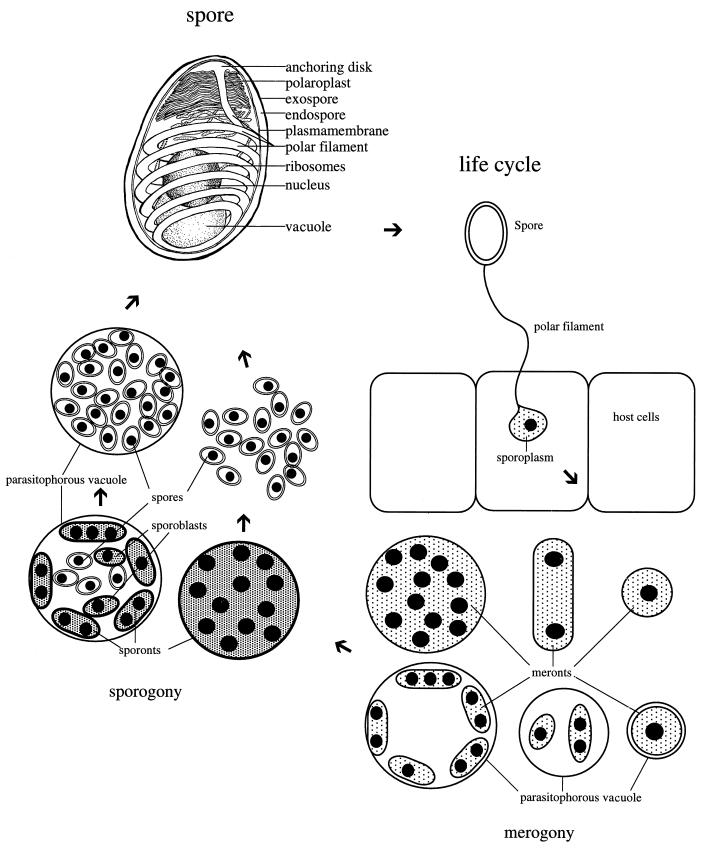
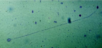
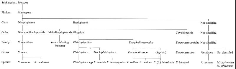
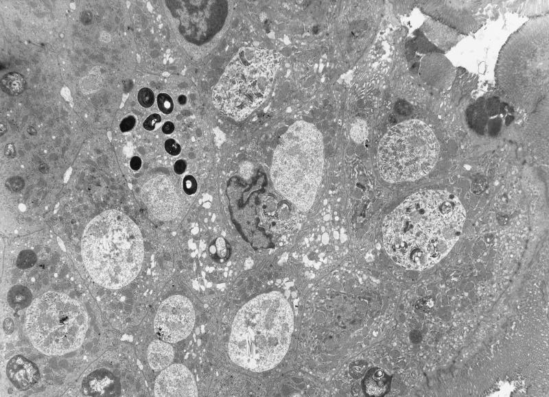
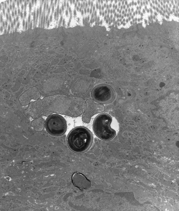
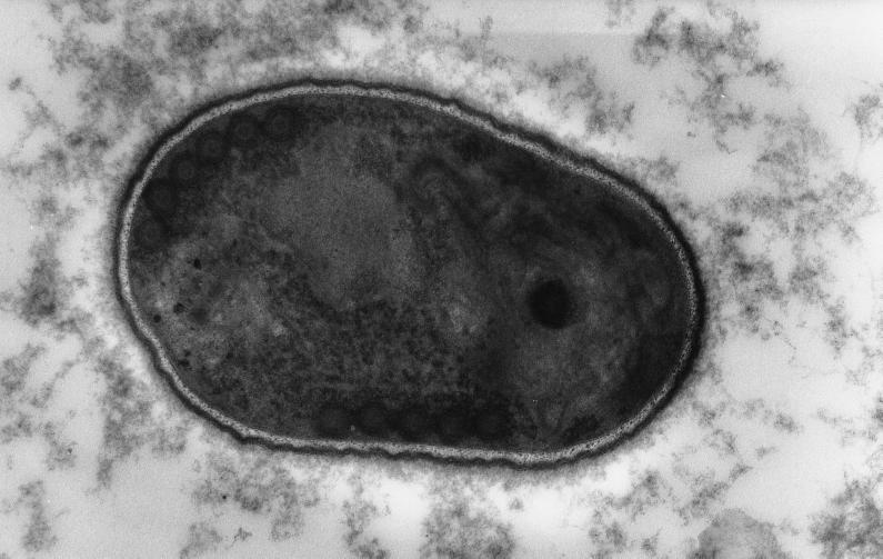
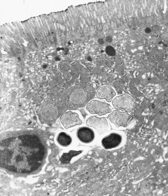
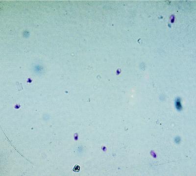
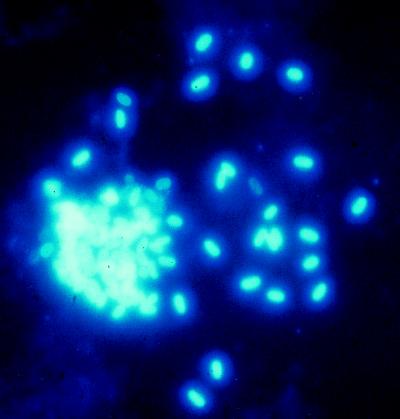
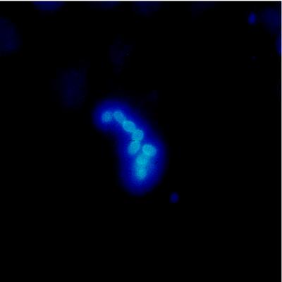
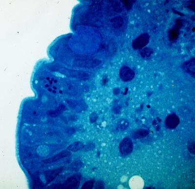
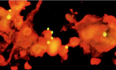
Microsporidia are obligate intracellular protozoan parasites that infect a broad range of vertebrates and invertebrates. These parasites are now recognized as one of the most common pathogens in human immunodeficiency virus-infected patients. For most patients with infectious diseases, microbiological isolation and identification techniques offer the most rapid and specific determination of the etiologic agent. This is not a suitable procedure for microsporidia, which are obligate intracellular parasites requiring cell culture systems for growth. Therefore, the diagnosis of microsporidiosis currently depends on morphological demonstration of the organisms themselves. Although the diagnosis of microsporidiosis and identification of microsporidia by light microscopy have greatly improved during the last few years, species differentiation by these techniques is usually impossible and transmission electron microscopy may be necessary. Immunfluorescent-staining techniques have been developed for species differentiation of microsporidia, but the antibodies used in these procedures are available only at research laboratories at present. During the last 10 years, the detection of infectious disease agents has begun to include the use of nucleic acid-based technologies. Diagnosis of infection caused by parasitic organisms is the last field of clinical microbiology to incorporate these techniques and molecular techniques (e.g., PCR and hybridization assays) have recently been developed for the detection, species differentiation, and phylogenetic analysis of microsporidia. In this paper we review human microsporidial infections and describe and discuss these newly developed molecular techniques.


Figures

FIG. 1
FIG. 1
Diagram of a microsporidian spore and representative life cycle (merogonic and sporogonic stages vary among different genera).
FIG. 2
FIG. 2
Giemsa stain of Nosema algerae spore with an extruded polar tube and with the sporoplasm at the end of the tube. Giemsa stain. Magnification, ×640.
FIG. 3
FIG. 3
Taxonomy of microsporidia infecting humans, using the revised taxonomy of Sprague et al. from 1992 (337), modified in light of the new taxonomic classification of Vittaformae corneae (324), Encephalitozoon intestinalis (10, 166), Trachipleistophora hominis (180), and T. antropophtera (356a). Comparing this taxonomic system with phylogenetic trees generated on the basis of DNA sequence data (see Fig. 13), it seems clear that the system of Sprague et al. (337) is flawed.
FIG. 4
FIG. 4
Transmission electron micrograph of duodenal epithelium from an HIV-infected patient heavily parasitized with Enterocytozoon bieneusi. Several cells are infected with merogonic and sporogonic stages, and one cell is infected with darkly staining spores. Magnification, ×6,600.
FIG. 5
FIG. 5
Transmission electron micrograph of duodenal epithelium from an HIV-infected patient infected with Encephalitozoon cuniculi. Two meronts with single nuclei, four sporonts with a thickened plasma membrane and highly developed endoplasmatic reticulum, and four spores inside a parasitophorous vacuole are visible. Magnification, ×17,500.
FIG. 6
FIG. 6
Transmission electron micrograph of an Encephalitozoon cuniculi spore in nasal discharge from a patient with AIDS and chronic rhinosinusitis. The spore contains a polar tubule with six coils lying in a single row and is coated with an electron-dense exospore. Magnification, ×55,125.
FIG. 7
FIG. 7
Transmission electron micrograph of duodenal epithelium of an HIV-infected patient infected with Encephalitozoon intestinalis. One meront with two nuclei, four sporonts with thickened plasma membrane, and four spores are separated by amorphous material which leads to septation of the parasitophorous vacuole. Magnification, ×17,500.
FIG. 8
FIG. 8
Encephalitozoon cuniculi spores in conjunctival swab from an HIV-infected patient with disseminated infection. Modified chromotrope-based stain. Magnification, ×870.
FIG. 9
FIG. 9
Encephalitozoon intestinalis spores derived from in vitro culture. Fluorescence microscopy after Uvitex 2B stain. Magnification, ×1,000.
FIG. 10
FIG. 10
Paraffin-embedded duodenal biopsy specimen from a patient with AIDS with intestinal Enterocytozoon bieneusi infection. The microsporidial spores are easily visualized within the enterocytes. Fluorescence microscopy after Uvitex 2B stain. Magnification, ×1,000.
FIG. 11
FIG. 11
Resin-embedded semithin (1-μm) section of duodenal mucosa from a patient with AIDS and intestinal Enterocytozoon bieneusi infection. Epithelial cells contain spores of Enterocytozoon bieneusi. Toluidine blue stain. Magnification, ×800.
FIG. 12
FIG. 12
Indirect immunofluorescent staining of Encephalitozoon cuniculi spores in nasal discharge of an HIV-infected patient with disseminated infection, using polyclonal anti-Encephalitozoon cuniculi antiserum. Magnification, ×400.
FIG. 13
FIG. 13
Tree representing the phylogenetic relationship of several microsporidian species as determined by SSU rRNA sequence analysis. This was the most pasimonious tree found by using the branch-and-bound option of PAUP. Reprinted from reference 1 with permission of the publisher.

References

    1. Aarons E J, Woodrow D, Hollister W S, Canning E U, Francis N, Gazzard B G. Reversible renal failure caused by a microsporidian infection. AIDS. 1994;8:1119–1121. - PubMed
    1. Albrecht H, Stellbrinck H J, Sobottka I. Failure of itraconazole to prevent Enterocytozoon bieneusi infection. Genitourin Med. 1995;71:325–326. - PMC - PubMed
    1. Albrecht H, Sobottka I, Stellbrinck H J, Greten H. Does choice of Pneumocystis carinii prophylaxis influence the prevalence of Enterocytozoon bieneusi microsporidiosis in AIDS patients? AIDS. 1995;9:302–304. - PubMed
    1. Aldras A M, Orenstein J M, Kotler D P, Shadduck J A, Didier E S. Detection of microsporidia by indirect immunofluorescence antibody test using polyclonal and monoclonal antibodies. J Clin Microbiol. 1994;32:608–612. - PMC - PubMed
    1. Anwar-Bruni D M, Hogan S E, Schwartz D A, Mel Wilcox C, Bryan R T, Lennox J L. Atovaquone is effective treatment for symptoms of gastrointestinal microsporidiosis in HIV-1-infected patients. AIDS. 1996;10:619–623. - PubMed

Publication types

LinkOut - more resources