. 1999 Apr;103(7):971-8.
doi: 10.1172/JCI5457.

Ordering of ceramide formation, caspase activation, and mitochondrial changes during CD95- and DNA damage-induced apoptosis

Affiliations

Ordering of ceramide formation, caspase activation, and mitochondrial changes during CD95- and DNA damage-induced apoptosis

A D Tepper et al. J Clin Invest. 1999 Apr.

Erratum in

  • J Clin Invest 1999 May;103(9):1363

Abstract

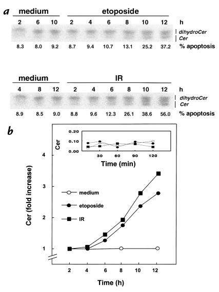
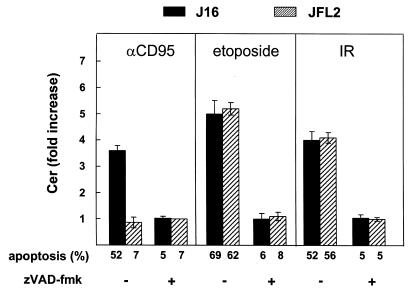
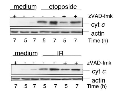
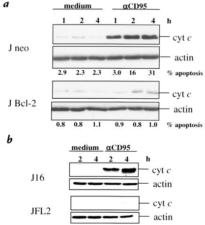
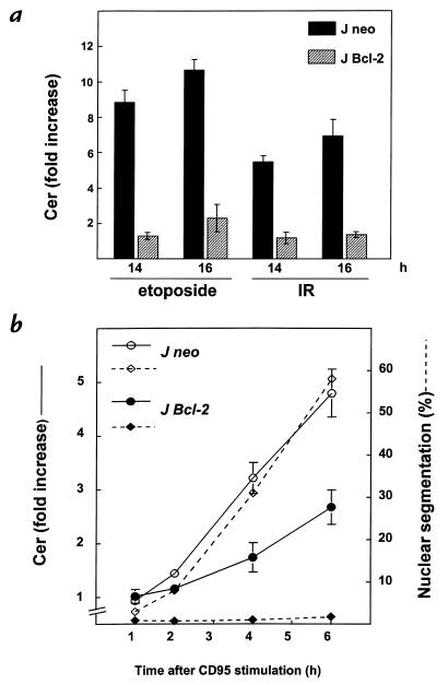
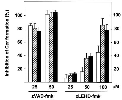
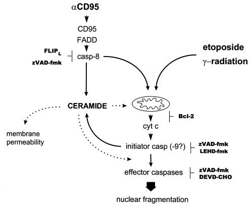
To evaluate the role of ceramide (Cer) in apoptosis signaling, we examined Cer formation induced by CD95, etoposide, or gamma-radiation (IR) in relation to caspase activation and mitochondrial changes in Jurkat T cells. The Cer response to all three stimuli was mapped in between caspases sensitive to benzoyloxycarbonyl-VAD-fluoromethylketone (zVAD-fmk) and acetyl-DEVD-aldehyde (DEVD-CHO). Cer production was independent of nuclear fragmentation but associated with the occurrence of other aspects of the apoptotic morphology. Caspase-8 inhibition abrogated Cer formation and apoptosis induced by CD95 but did not affect the response to etoposide or IR, placing CD95-induced Cer formation downstream from caspase-8 and excluding a role for caspase-8 in the DNA damage pathways. CD95 signaling to the mitochondria required caspase-8, whereas cytochrome c release in response to DNA damage was caspase-independent. These results indicate that the caspases required for the Cer response to etoposide and IR reside at or downstream from the mitochondria. Bcl-2 overexpression abrogated the Cer response to etoposide and IR and reduced CD95-induced Cer accumulation. We conclude that the Cer response to DNA damage fully depends on mitochondrion-dependent caspases, whereas the response to CD95 partially relies on these caspases. Our data imply that Cer is not instrumental in the activation of inducer caspases or signaling to the mitochondria. Rather, Cer formation is associated with the execution phase of apoptosis.


Figures

Figure 1
Figure 1
Etoposide and IR induce Cer formation and apoptosis. Jurkat T cells (J16) labeled until equilibrium with [14C]serine were exposed to etoposide (10 μg/ml) or IR (30 Gy), or left untreated (medium). At the indicated times, Cer levels and apoptosis were determined. (a) TLC separation of Cer from dihydroceramide. Apoptosis is expressed as percentage of nuclei with subdiploid DNA content. (b) Time course of changes in Cer levels induced by etoposide or IR. Cer data are expressed as -fold increase relative to control and are representative of three experiments. The inset shows Cer levels (expressed relative to total radioactivity in phosphatidylserine and phosphatidylethanolamine) at shorter time points. Cer, ceramide; IR, γ-radiation.
Figure 2
Figure 2
Role of caspases in CD95-, etoposide-, and IR-induced Cer accumulation and apoptosis. Control Jurkat cells (J16; filled bars) or Jurkat cells stably transfected with FLIPL cDNA (JFL2; hatched bars) (34) were treated with anti-CD95 mAb (200 ng/ml) for 6 h, or etoposide (10 μg/ml) or IR (30 Gy) for 16 h. Where indicated (+), cells were pretreated (2 h) and further incubated with 50 μM zVAD-fmk. Cer levels were quantitated and expressed as -fold increase relative to time-matched medium controls. Apoptosis sensitivity of J16 and JFL2 was determined in parallel and expressed as percentage hypoploid cells. Cer data are representative of two independent experiments and show mean ± SD from duplicate samples in one experiment. FLIPL, FLICE-inhibitory protein; mAb, monoclonal antibody; zVAD-fmk, benzoyloxycarbonyl-VAD-fluoromethylketone.
Figure 3
Figure 3
Etoposide and IR induce cyt c release in a caspase-independent manner. Jurkat cells (J16) were treated with zVAD-fmk (2 h, 50 μM) or left untreated and then induced to undergo apoptosis by etoposide (10 μg/ml) or IR (30 Gy). Cytosolic extracts were prepared at the indicated times and separated by 12% SDS-PAGE. Cyt c and actin content were evaluated by immunoblot analysis. Cyt c, cytochrome c.
Figure 4
Figure 4
CD95 induces cyt c release in a Bcl-2– and FLIPL-inhibitable manner. Cytosolic extracts (10 μg of protein) of J neo and J Bcl-2 (a), and J16 and JFL2 (b), prepared after the indicated times of treatment with anti-CD95 mAb (200 ng/ml), were separated by SDS-PAGE, and cyt c and actin content were evaluated by immunoblotting.
Figure 5
Figure 5
Bcl-2 abrogates Cer formation induced by etoposide or IR and reduces CD95-induced Cer formation. (a) Jurkat cells transfected with vector (J neo; filled bars) or with human bcl-2 cDNA (J Bcl-2; hatched bars) labeled with [14C]serine were exposed to etoposide (10 μg/ml) or IR (30 Gy). Cer levels were determined after 14 and 18 h and expressed as -fold increase (means ± SD) relative to Cer levels in time-matched untreated cells. (b) J Neo or J Bcl-2 cells labeled with [14C]serine were treated with anti-CD95 mAb (200 ng/ml). Cer formation and nuclear segmentation were determined in the same set of samples at the indicated times. Solid lines show Cer expressed as -fold increase relative to control cells and represent three independent experiments. Dotted lines show the percentage of nuclear segmentation.
Figure 6
Figure 6
Differential effects of zVAD-fmk and zLEHD-fmk on Cer production induced by CD95 ligation or DNA damage. Jurkat cells (J16) labeled with [14C]serine were left untreated or preincubated for 2 h in the presence of zVAD-fmk or zLEHD-fmk at the indicated concentrations before addition of anti-CD95 mAb (200 ng/ml), etoposide (10 μg/ml), or exposure to IR (30 Gy). Quantitation of cellular Cer was performed after 5 h (anti-CD95; open bars) or 15 h (Eto/IR; hatched bars and filled bars, respectively) of further incubation. Results are expressed as percentage inhibition (means ± SD) of the Cer response observed in the absence of inhibitor peptide. Shown are data from a representative experiment containing duplicate samples performed two times and showing similar results. zLEHD-fmk, benzoyloxycarbonyl-LEHD-fluoromethylketone.
Figure 7
Figure 7
Mapping the Cer response in relation to caspase activation and mitochondrial changes in the CD95, etoposide, and IR pathways in Jurkat cells. Cer is placed in between initiator and executioner caspases in all cases, because its accumulation is inhibited by zVAD-fmk but not by DEVD-CHO. CD95 requires casp-8, whereas DNA damage–induced Cer formation does not involve casp-8 or death receptor signaling. In the case of etoposide or IR, the most proximal zVAD-fmk target resides at, or downstream from, the mitochondria, since zVAD-fmk does not block cyt c release. Bcl-2 overexpression abrogates Cer formation in response to etoposide or IR but only partially interferes with the CD95-induced Cer response. Possible roles of Cer are indicated by dotted lines. DEVD-CHO, acetyl-DEVD-aldehyde.

References

    1. Hannun YA. Functions of ceramide in coordinating cellular responses to stress. Science. 1996;274:1855–1859. - PubMed
    1. Wiegmann K, Schutze S, Machleidt T, Witte D, Kronke M. Functional dichotomy of neutral and acidic sphingomyelinases in tumor necrosis factor signaling. Cell. 1994;78:1005–1015. - PubMed
    1. Cifone MG, et al. Multiple pathways originate at the Fas/APO-1 (CD95) receptor: sequential involvement of phosphatidylcholine-specific phospholipase C and acidic sphingomyelinase in the propagation of the apoptotic signal. EMBO J. 1995;14:5859–5868. - PMC - PubMed
    1. Tepper CG, et al. Role for ceramide as an endogenous mediator of Fas-induced cytotoxicity. Proc Natl Acad Sci USA. 1995;92:8443–8447. - PMC - PubMed
    1. Santana P, et al. Acid sphingomyelinase-deficient human lymphoblasts and mice are defective in radiation-induced apoptosis. Cell. 1996;86:189–199. - PubMed

Publication types

MeSH terms