. 1999 Apr;103(7):1015-21.
doi: 10.1172/JCI5849.

Surfactant protein-A enhances respiratory syncytial virus clearance in vivo

Affiliations

Surfactant protein-A enhances respiratory syncytial virus clearance in vivo

A M LeVine et al. J Clin Invest. 1999 Apr.

Abstract

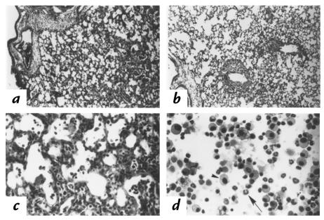
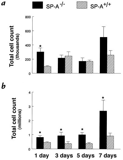
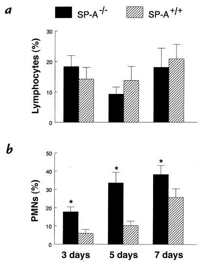
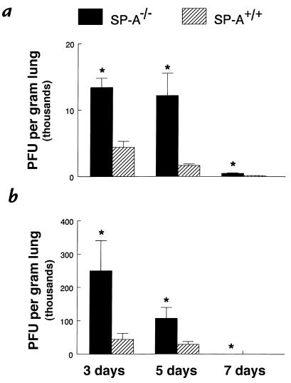
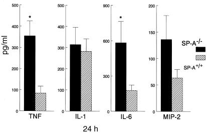
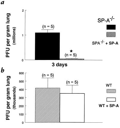
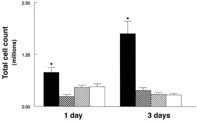
To determine the role of surfactant protein-A(SP-A) in antiviral host defense, mice lacking SP-A (SP-A-/-) were produced by targeted gene inactivation. SP-A-/- and control mice (SP-A+/+) were infected with respiratory syncytial virus (RSV) by intratracheal instillation. Pulmonary infiltration after infection was more severe in SP-A-/- than in SP-A+/+ mice and was associated with increased RSV plaque-forming units in lung homogenates. Pulmonary infiltration with polymorphonuclear leukocytes was greater in the SP-A-/- mice. Levels of proinflammatory cytokines tumor necrosis factor-alpha and interleukin-6 were enhanced in lungs of SP-A-/- mice. After RSV infection, superoxide and hydrogen peroxide generation was deficient in macrophages from SP-A-/- mice, demonstrating a critical role of SP-A in oxidant production associated with RSV infection. Coadministration of RSV with exogenous SP-A reduced viral titers and inflammatory cells in the lung of SP-A-/- mice. These findings demonstrate that SP-A plays an important host defense role against RSV in vivo.


Figures

Figure 1
Figure 1
Pulmonary pathology after RSV infection. Photomicrographs of lung parenchyma in (a) SP-A–/– and (b) SP-A+/+ mice were prepared 7 days after intratracheal administration of 107 pfu RSV. Histological sections were stained with hematoxylin and eosin. Infiltration was observed surrounding the terminal bronchi and pulmonary vessels in the SP-A–/– and SP-A+/+ mice. Higher magnification demonstrates infiltration with mononuclear cells and polymorphonuclear leukocytes in SP-A–/– mice (c). Cells from BAL consisted of mononuclear cells (arrowhead) and polymorphonuclear leukocytes (arrow) (d). a and b: ×10; c and d: ×40. BAL, bronchoalveolar lavage; pfu, plaque-forming units; RSV, respiratory syncytial virus; SP-A, surfactant protein-A.
Figure 2
Figure 2
Increased total cell counts in BAL fluid from SP-A–/– mice. Lung cells were recovered by BAL, stained with trypan blue, and counted under light microscopy. (a) The low dose of RSV (106 pfu) caused less migration of inflammatory cells in the lungs from both genotypes; however, an earlier influx of cells was found in SP-A–/– (filled bars) BAL fluid than in SP-A+/+ (hatched bars) mice. (b) SP-A–/– mice infected with 107 pfu RSV had significantly greater total cell counts in BAL fluid at all time points. Data are mean ± SEM with n = 5 mice per group (a) and n = 9 mice per group (b). *P < 0.05 compared with SP-A+/+ mice.
Figure 3
Figure 3
Increased neutrophils in BAL fluid from SP-A–/– mice after RSV infection. Cytospin preparations of BAL fluid were stained with Diff-Quik to identify macrophages, lymphocytes, and polymorphonuclear leukocytes. (a) The percentage of neutrophils in BAL fluid was significantly greater 3, 5, and 7 days after administration of 107 pfu RSV to SP-A–/– (filled bars) compared with SP-A+/+ (hatched bars) mice. (b) Percentages of lymphocytes in BAL fluid were not different between the two groups. Data are mean ± SEM with n = 9 mice per group. *P < 0.05 compared with SP-A+/+ mice. PMNs, polymorphonuclear leukocytes.
Figure 4
Figure 4
Increased RSV in lung homogenates from SP-A–/– mice. RSV titers were determined by quantitative plaque assays of lung homogenates. Viral titers were significantly greater 3, 5, and 7 days after administration of both 106 (a) and 107 (b) pfu RSV in SP-A–/– (filled bars) compared with SP-A+/+ (hatched bars) mice. Data are mean ± SEM with n = 6 mice per group (a) and n = 10 mice per group (b). *P < 0.05 compared with SP-A+/+ mice.
Figure 5
Figure 5
Increased tumor necrosis factor-α (TNF) and interleukin-6 (IL-6) in lung homogenates from SP-A–/– mice after RSV infection. Concentrations of TNF-α, interleukin-1β (IL-1), IL-6, and macrophage inflammatory protein-2 (MIP-2) were assessed in lung homogenates from SP-A–/– (filled bars) and SP-A+/+ (hatched bars) mice. Increased concentrations of TNF-α and IL-6 were found in lung homogenates from the SP-A–/– mice at 24 h. Although macrophage inflammatory MIP-2 was increased in SP-A–/– at 24 h, differences did not reach statistical significance. Data are expressed as picograms per milliliter and represent mean ± SEM with n = 10 mice per group. *P < 0.05 compared with SP-A+/+ mice.
Figure 6
Figure 6
SP-A enhanced RSV clearance from the lung of SP-A–/– mice. RSV titers were determined by quantitative plaque assays of lung homogenates 3 days after infection with 107 pfu of RSV. Viral titers in the lung were significantly reduced in SP-A–/– mice treated with SP-A (100 μg) (cross-hatched bar) compared with untreated SP-A–/– mice (filled bar) (a). Treatment of SP-A–/– mice with SP-A reduced viral titers to the wild-type level. Viral titers were similar in SP-A–treated (open bar) and untreated wild-type mice (hatched bar) (b). Data are mean ± SEM. *P < 0.05 compared with SP-A–/– mice. WT, wild-type.
Figure 7
Figure 7
SP-A decreased inflammatory cells in BAL fluid from SP-A–/– mice. After RSV infection (107 pfu), lung cells were recovered by BAL, stained with trypan blue, and counted under light microscopy. One and 3 days after RSV infection, total cell counts in BAL fluid were significantly reduced in SP-A–/– mice treated with SP-A (100 μg) (cross-hatched bars) compared with untreated SP-A–/– mice (filled bars). Treatment of SP-A–/– mice reduced BAL cell counts to the wild-type level (hatched bars). Total cell counts in BAL fluid were similar in SP-A–treated (open bars) and untreated wild-type mice (hatched bars). Data are mean ± SEM. *P < 0.05 compared with SP-A+/+ mice.

References

    1. Thiel S, Reid K. Structures and functions associated with the group of mammalian lectins containing collagen-like sequences. FEBS Lett. 1989;250:78–84. - PubMed
    1. Sastry K, Ezekowitz RA. Collectins: pattern recognition molecules involved in first line host defense. Curr Opin Immunol. 1993;5:59–66. - PubMed
    1. Super M, Thiel S, Lu J, Levinsky RJ, Turner MW. Association of low levels of mannan-binding protein with a common defect of opsonisation. Lancet. 1989;2:1236–1239. - PubMed
    1. Malhotra R, Thiel S, Reid KB, Sim RB. Human leukocyte C1q receptor binds other soluble proteins with collagen domains. J Exp Med. 1990;172:955–959. - PMC - PubMed
    1. Friis-Christiansen P, et al. In vivo and in vitro antibacterial activity of conglutinin, a mammalian plasma lectin. Scand J Immunol. 1990;31:453–460. - PubMed

Publication types

MeSH terms