. 1999 Apr;103(7):1039-46.
doi: 10.1172/JCI5779.

Proteolysis of monocyte CD14 by human leukocyte elastase inhibits lipopolysaccharide-mediated cell activation

Affiliations

Proteolysis of monocyte CD14 by human leukocyte elastase inhibits lipopolysaccharide-mediated cell activation

K Le-Barillec et al. J Clin Invest. 1999 Apr.

Abstract

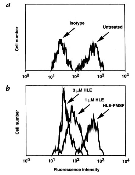
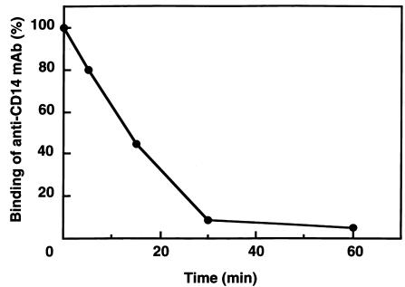
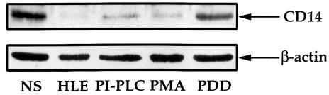
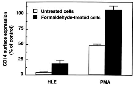
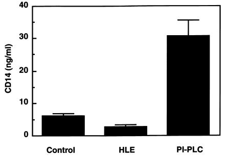
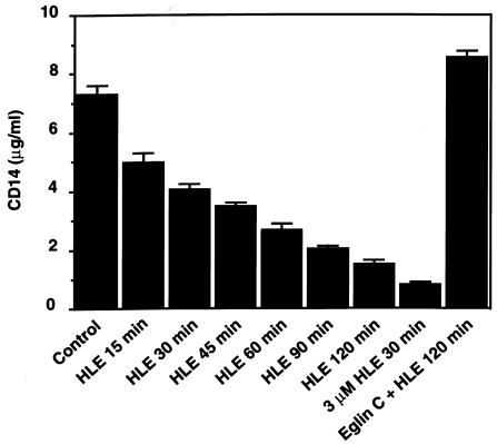
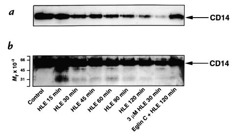
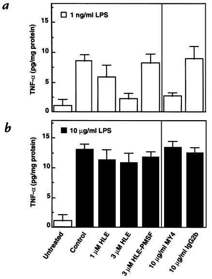
Human leukocyte elastase (HLE), a polymorphonuclear neutrophil (PMN) serine proteinase, is proteolytically active on some membrane receptors at the surface of immune cells. The present study focused on the effect of HLE on the expression of CD14, the main bacterial lipopolysaccharide (LPS) receptor at the surface of monocytes. HLE exhibited a time- and concentration-dependent downregulatory effect on CD14 surface expression. A 30-minute incubation of 3 microM HLE was required to display 95% disappearance of the receptor. This downregulation resulted from a direct proteolytic process, not from a shedding consecutive to monocyte activation as observed upon challenge with phorbol myristate acetate (PMA). To confirm that CD14 is a substrate for HLE, this enzyme was incubated with recombinant human CD14 (Mr approximately 57,000), and proteolysis was further analyzed by immunoblot analysis. Cleavage of the CD14 molecule was directly evidenced by the generation of short-lived fragments (Mr approximately 47,000 and 30,000). As a consequence of the CD14 proteolysis, a decrease in the responsiveness of monocytes to LPS was observed, as assessed by measuring tumor necrosis factor-alpha (TNF-alpha) formation. This inhibition was only observed with 1 ng/ml of LPS, i.e., when only the CD14-dependent pathway was involved. At a higher LPS concentration, such as 10 microgram/ml, when CD14-independent pathways were operative, this inhibition was overcome. The direct proteolysis by HLE of the membrane CD14 expressed on monocytes illustrates a potential anti-inflammatory effect of HLE through inhibition of LPS-mediated cell activation.


Figures

Figure 1
Figure 1
Effect of HLE on the surface expression of CD14 on human monocytes. Monocytes were treated with 1 or 3 μM HLE or 3 μM HLE-PMSF for 30 min at 37°C, and reactions were stopped as described in Methods. Binding of the MY4 antibody or a matched isotype was then analyzed by flow cytometry. (a) Nontreated cells were labeled with the MY4 antibody (Untreated) or a matched IgG2b (Isotype). (b) Treated cells (as indicated) were labeled with MY4 antibody. Fluorescence tracings are representative of three distinct experiments. HLE, human leukocyte elastase.
Figure 2
Figure 2
Time course of reduction by HLE of the surface expression of CD14 on human monocytes. Monocytes were treated with 3 μM HLE for various time periods at 37°C. CD14 expression was analyzed by flow cytometry using the MY4 antibody. The binding of MY4 is expressed as the percentage of the control value obtained with nontreated cells. Results represent one experiment out of two obtained from different donors.
Figure 3
Figure 3
Immunoblot analysis of CD14 expression by human monocytes. Monocytes were untreated (NS) or treated with 3 μM HLE, 0.6 U/ml PI-PLC, 15 nM PMA, or 15 nM PDD for 30 min at 37°C. Reactions were stopped with 20 μM eglin C, and monocytes were immediately solubilized with Triton/SDS. After SDS-PAGE on 10% acrylamide gels and transfer to nitrocellulose membranes, samples were probed with a polyclonal anti-CD14 antibody. In addition, the same samples were probed with an anti–β-actin antibody to control the equal loading of the different wells. Relative molecular mass was calculated with respect to calibration-standard proteins included in the gel. PDD, phorbol 12,13-didecanoate; PI-PLC, phosphatidylinositol-phospholipase C; PMA, phorbol myristate acetate.
Figure 4
Figure 4
Effect of HLE on the modulation of CD14 surface expression after formaldehyde treatment of monocytes. Monocytes were fixed or not with 1% formaldehyde for 10 min at room temperature, and the fixation process was neutralized with 12 mM NH4Cl. After washing, cells were incubated with 3 μM HLE or 15 nM PMA for 30 min at 37°C; at the end of the reaction, 20 μM eglin C was added. Binding of the MY4 anti-CD14 antibody is expressed as the percentage of control values obtained with nontreated cells. Results are means ± SEM of three experiments.
Figure 5
Figure 5
Measurements of CD14 in the extracellular medium of HLE-treated monocytes. Monocytes were treated with 3 μM HLE or 0.6 U/ml PI-PLC for 30 min at 37°C; at the end of the reaction, 20 μM eglin C was added. The extracellular medium was then analyzed for CD14 by ELISA. Data are means ± SEM of four to seven experiments with different donors.
Figure 6
Figure 6
Time course of the disappearance of recombinant CD14 upon incubation with HLE. Recombinant CD14 (0.25 μM) was incubated with 500 nM or 3 μM (where indicated) HLE for various time periods at 37°C. A mixture of 20 μM eglin C and 500 nM HLE was incubated for 2 h under the same conditions. Reactions were stopped with 20 μM eglin C, and samples were next analyzed for CD14 by ELISA. Data are means ± SEM of three distinct experiments.
Figure 7
Figure 7
Immunoblot analysis of the time course of HLE-treated recombinant CD14. Recombinant CD14 (0.25 μM) was incubated with 500 nM or 3 μM (where indicated) HLE for various time periods at 37°C. A mixture of 20 μM eglin C and 500 nM HLE was incubated for 2 h under the same conditions. Reactions were stopped with 20 μM eglin C, and samples were processed for CD14 immunoblot analysis using a 15% polyacrylamide gel. Relative molecular masses were calculated with respect to calibration-standard proteins included in the gel. (a) Membrane exposed for 1 min; (b) same membrane exposed for 2 h.
Figure 8
Figure 8
Inhibition by HLE of TNF-α synthesis by LPS-activated human monocytes. Monocytes were preincubated as indicated under each column for 30 min at 37°C, and reactions were stopped by 20 μM eglin C. Cells were then washed twice with medium before stimulation with (a) 1 ng/ml or (b) 10 μg/ml LPS for 3 h at 37°C. Supernatants were analyzed for TNF-α content by ELISA. Data represent means ± SEM of three experiments with cells from different donors. LPS, lipopolysaccharide; TNF-α, tumor necrosis factor-α.

References

    1. Smith JA. Neutrophils, host defense, and inflammation: a double-edged sword. J Leukoc Biol. 1994;56:672–686. - PubMed
    1. Woessner, J.F., Jr. 1992. Role of cellular proteinases and their protein inhibitors in inflammation. In Biochemistry of inflammation. J.T. Whicher and S.W. Evans, editors. Kluwer Academic Publishers. Lancaster, PA. 57–89.
    1. Döring G. The role of neutrophil elastase in chronic inflammation. Am J Respir Crit Care Med. 1994;150:S114–S117. - PubMed
    1. Travis J, Pike R, Imamura T, Potempa J. The role of proteolytic enzymes in the development of pulmonary emphysema and periodontal disease. Am J Respir Crit Care Med. 1994;150:S143–S146. - PubMed
    1. Bieth, J.G. 1986. Elastases: catalytic and biological properties. In Regulation of matrix accumulation. R. Mecham, editor. Academic Press. New York, NY. 217–230.

Publication types