. 1999 Apr;103(7):1067-76.
doi: 10.1172/JCI6246.

Disruption of LDL receptor gene in transgenic SREBP-1a mice unmasks hyperlipidemia resulting from production of lipid-rich VLDL

Affiliations

Disruption of LDL receptor gene in transgenic SREBP-1a mice unmasks hyperlipidemia resulting from production of lipid-rich VLDL

J D Horton et al. J Clin Invest. 1999 Apr.

Abstract

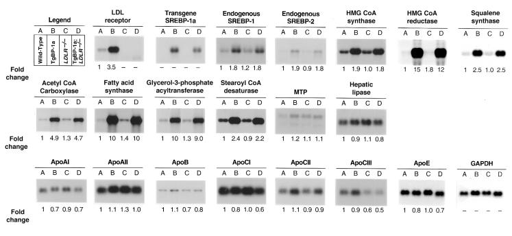
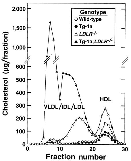
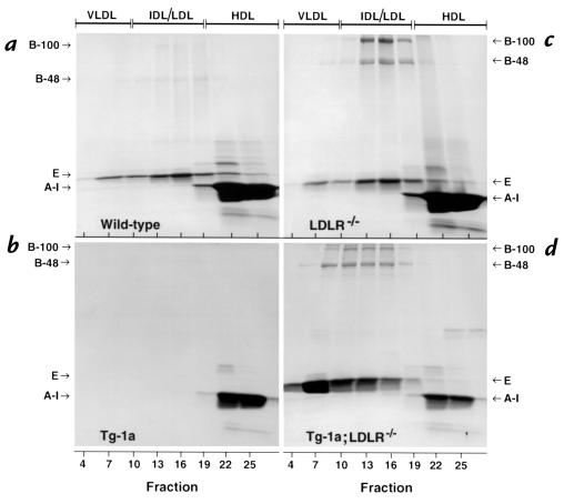
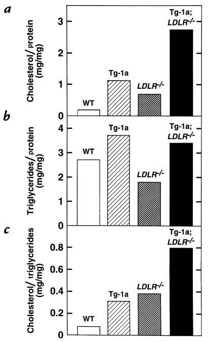
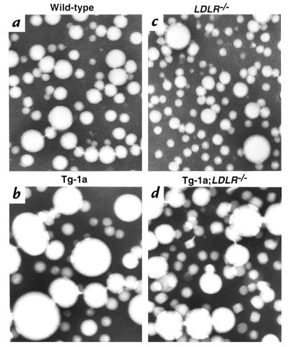
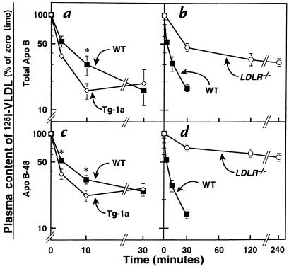
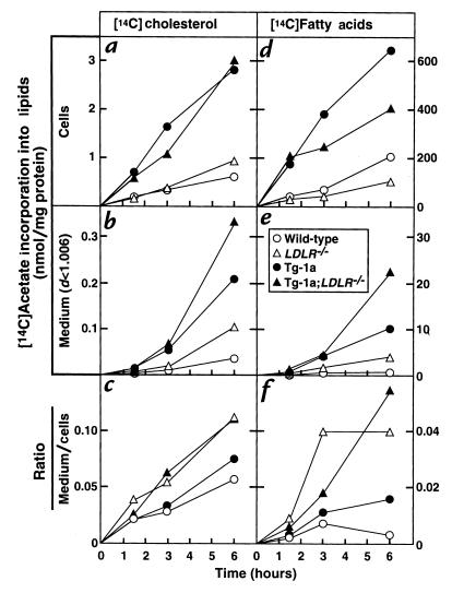
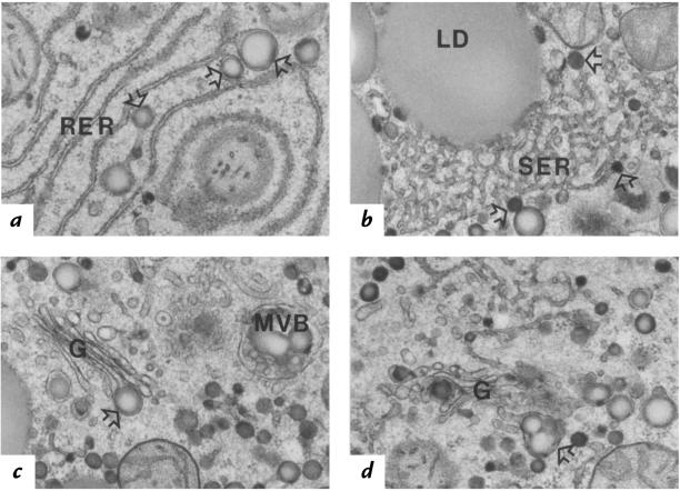
Transgenic mice that overexpress the nuclear form of sterol regulatory element binding protein-1a (SREBP-1a) in liver (TgBP-1a mice) were shown previously to overproduce cholesterol and fatty acids and to accumulate massive amounts of cholesterol and triglycerides in hepatocytes. Despite the hepatic overproduction of lipids, the plasma levels of cholesterol ( approximately 45 mg/dl) and triglycerides ( approximately 55 mg/dl) were not elevated, perhaps owing to degradation of lipid-enriched particles by low-density lipoprotein (LDL) receptors. To test this hypothesis, in the current studies we bred TgBP-1a mice with LDL receptor knockout mice. As reported previously, LDLR-/- mice manifested a moderate elevation in plasma cholesterol ( approximately 215 mg/dl) and triglycerides ( approximately 155 mg/dl). In contrast, the doubly mutant TgBP-1a;LDLR-/- mice exhibited marked increases in plasma cholesterol ( approximately 1,050 mg/dl) and triglycerides ( approximately 900 mg/dl). These lipids were contained predominantly within large very-low-density lipoprotein (VLDL) particles that were relatively enriched in cholesterol and apolipoprotein E. Freshly isolated hepatocytes from TgBP-1a and TgBP-1a;LDLR-/- mice overproduced cholesterol and fatty acids and secreted increased amounts of these lipids into the medium. Electron micrographs of livers from TgBP-1a mice showed large amounts of enlarged lipoproteins within the secretory pathway. We conclude that the TgBP-1a mice produce large lipid-rich lipoproteins, but these particles do not accumulate in plasma because they are degraded through the action of LDL receptors.


Figures

Figure 1
Figure 1
Amounts of various mRNAs in livers of (A) wild-type, (B) TgBP-1a, (C) LDLR–/–, and (D) TgBP-1a;LDLR–/– mice, as measured by blot hybridization. Total RNA isolated from livers of mice described in experiment A of Table 1 was pooled, and 15-μg aliquots were subjected to electrophoresis and blot hybridization with the indicated 32P-labeled probe. The amount of radioactivity in each band was quantified as described in Methods. The probe used for SCD was a mouse SCD-1 cDNA fragment that detects both SCD-1 and SCD-2 mRNAs (see ref. 35). The increase for each mRNA relative to that of wild-type mice was calculated after correction for loading differences with GAPDH as described previously (12). These values are shown below each blot. GAPDH, glyceraldehyde-3-phosphate dehydrogenase; MTP, microsomal triglyceride transfer protein; SCD, stearoyl CoA desaturase.
Figure 2
Figure 2
FPLC profiles of plasma lipoproteins from wild-type (open circles), TgBP-1a (filled circles), LDLR–/– (open triangles), and TgBP-1a;LDLR–/– (filled triangles) mice. Plasma from mice described in experiment B of Table 1 was pooled (2.5 ml for each genotype) and subjected to ultracentrifugation at d = 1.215 g/ml. The lipoprotein fractions (d < 1.215 g/ml) were subjected to gel filtration by FPLC, and the cholesterol content of each fraction was measured as described in Methods. FPLC, fast performance liquid chromatography.
Figure 3
Figure 3
SDS-PAGE of plasma apolipoproteins from (a) wild-type, (b) TgBP-1a, (c) LDLR–/–, and (d) TgBP-1a;LDLR–/– mice. Equal aliquots (0.75 ml) from three consecutive FPLC fractions shown in Figure 2 were pooled (total of 1.5 ml) and delipidated, and the apoproteins were precipitated as described in Methods. The apoproteins were subjected to 3–15% gradient SDS-PAGE and were stained with Coomassie blue. The positions of migration of apo B-100, apo B-48, apo E, and apo AI are indicated.
Figure 4
Figure 4
Composition of plasma VLDL (d < 1.006 g/ml) from wild-type (WT), TgBP-1a, LDLR–/–, and TgBP-1a;LDLR–/– mice. The ratios of cholesterol/protein, triglycerides/protein, and cholesterol/triglycerides were calculated from the values shown in Table 2. VLDL, very-low-density lipoprotein.
Figure 5
Figure 5
Electron microscopy of negatively stained plasma VLDL (d < 1.006 g/ml) from (a) wild-type, (b) TgBP-1a, (c) LDLR–/–, and (d) TgBP-1a;LDLR–/–mice. After a 4-h fast, plasma samples from four male mice of each genotype were pooled, and VLDL was isolated for viewing by electron microscopy as described in Methods. ×108,000.
Figure 6
Figure 6
Plasma clearance of 125I-labeled VLDL in wild-type (WT), TgBP-1a, LDLR–/–, and TgBP- 1a;LDLR–/– mice. VLDL was isolated from four fasted male TgBP-1a;LDLR–/– mice and labeled with 125I as described in Methods. 125I-labeled VLDL (15 μg protein, 860 cpm/ng apo B protein) was injected intravenously into each of five 12-week-old male mice with the indicated genotype. Blood was drawn at the indicated times to quantify the content of 125I-labeled apo B in plasma. (a and b) Plasma content of 125I-labeled total apo B was measured by isopropanol precipitation followed by scintillation counting (as described in Methods). (c and d) Plasma samples were subjected to SDS-PAGE, and the signal generated by apo B-48 was quantified using a Fuji PhosphorImager as described in Methods. The data are plotted as the percent of the zero time value. *P < 0.05 (Student’s t test) compared with wild-type values. Each value represents the mean ± SEM of data from five mice.
Figure 7
Figure 7
Rates of lipid synthesis and secretion by primary hepatocytes from wild-type (open circles), TgBP-1a (filled circles), LDLR–/– (open triangles), and TgBP-1a;LDLR–/– (filled triangles) mice. Primary hepatocytes were prepared from one mouse of each genotype as described in Methods. The hepatocytes were incubated with DMEM containing 5% FCS for 3 h at 37°C, after which the cells were incubated with 0.5 mM sodium [14C]acetate (18 dpm/pmol) in DMEM supplemented with 5% human lipoprotein–deficient serum. At the indicated time, the medium was removed for centrifugation at d < 1.006 g/ml, the monolayers were washed, and the cells were harvested. The content of 14C-labeled cholesterol and fatty acids in cells and medium was quantified as described in Methods. Each value is the average of duplicate incubations. Similar results were obtained in three other experiments in which different groups of mice were studied at a single time point.
Figure 8
Figure 8
Electron micrographs of thin sections depicting lipoprotein assembly and degradation in fatty hepatocytes of TgBP-1a mouse liver. These hepatocytes contained numerous very large (∼100–250 nm) nascent VLDL particles (open arrows) in the following areas: (a) within the lumen of the unique smooth-surfaced (ribosome-free) terminal ends of rough ER (RER); (b) within the smooth ER (SER) proper; (c and d) within forming secretory vesicles of the Golgi apparatus (G), singly (c) or clustered (d). Multivesicular bodies (MVB) that contained partially degraded lipoproteins the same size as nascent VLDL were often seen (c). Larger than normal patches of tightly packed membranes of SER were common in these fatty hepatocytes and sometimes appeared to be associated with cytosolic lipid droplets (LD) (b). Differences in the intensity of lipid staining between nascent VLDL particles of different diameters may reflect differential absorbency of the imidazole mordant and/or differences in unsaturated fatty acid content of the core lipids (27). ×27,000.

References

    1. Brown MS, Goldstein JL. The SREBP pathway: regulation of cholesterol metabolism by proteolysis of a membrane-bound transcription factor. Cell. 1997;89:331–340. - PubMed
    1. Hua X, Sakai J, Ho YK, Goldstein JL, Brown MS. Hairpin orientation of sterol regulatory element binding protein-2 in cell membranes as determined by protease protection. J Biol Chem. 1995;270:29422–29427. - PubMed
    1. Tontonoz P, Kim JB, Graves RA, Spiegelman BM. ADD1: a novel helix-loop-helix transcription factor associated with adipocyte determination and differentiation. Mol Cell Biol. 1993;13:4753–4759. - PMC - PubMed
    1. Shimomura I, Shimano H, Horton JD, Goldstein JL, Brown MS. Differential expression of exons 1a and 1c in mRNAs for sterol regulatory element binding protein-1 in human and mouse organs and cultured cells. J Clin Invest. 1997;99:838–845. - PMC - PubMed
    1. Hua X, Wu J, Goldstein JL, Brown MS, Hobbs HH. Structure of human gene encoding sterol regulatory element binding protein-1 (SREBF1) and localization of SREBF1 and SREBF2 to chromosomes 17p11.2 and 22q13. Genomics. 1995;25:667–673. - PubMed

Publication types

MeSH terms