. 1999 Apr 16;274(16):11139-49.
doi: 10.1074/jbc.274.16.11139.

A phosphotransferase that generates phosphatidylinositol 4-phosphate (PtdIns-4-P) from phosphatidylinositol and lipid A in Rhizobium leguminosarum. A membrane-bound enzyme linking lipid a and ptdins-4-p biosynthesis

Affiliations

A phosphotransferase that generates phosphatidylinositol 4-phosphate (PtdIns-4-P) from phosphatidylinositol and lipid A in Rhizobium leguminosarum. A membrane-bound enzyme linking lipid a and ptdins-4-p biosynthesis

S S Basu et al. J Biol Chem. .

Abstract

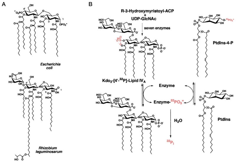
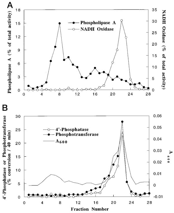
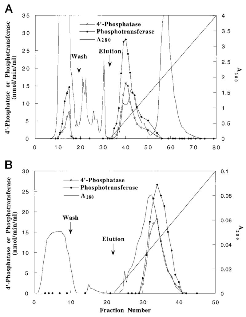
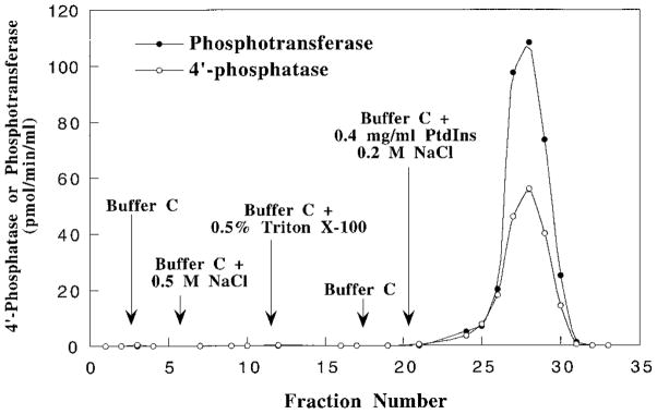
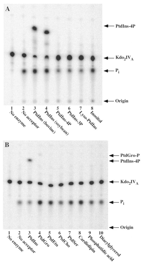
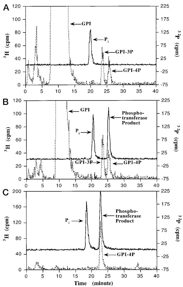
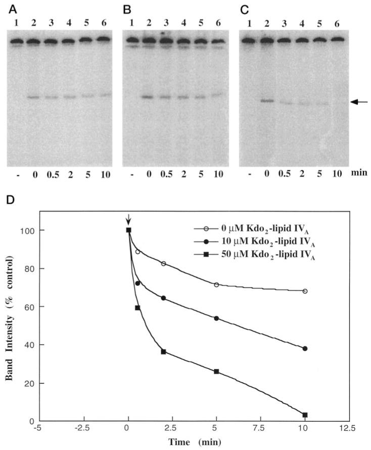
Membranes of Rhizobium leguminosarum contain a 3-deoxy-D-manno-octulosonic acid (Kdo)-activated lipid A 4'-phosphatase required for generating the unusual phosphate-deficient lipid A found in this organism. The enzyme has been solubilized with Triton X-100 and purified 80-fold. As shown by co-purification and thermal inactivation studies, the 4'-phosphatase catalyzes not only the hydrolysis of (Kdo)2-[4'-32P]lipid IVA but also the transfer the 4'-phosphate of Kdo2-[4'-32P]lipid IVA to the inositol headgroup of phosphatidylinositol (PtdIns) to generate PtdIns-4-P. Like the 4'-phosphatase, the phosphotransferase activity is not present in Escherichia coli, Rhizobium meliloti, or the nodulation-defective mutant 24AR of R. leguminosarum. The specific activity for the phosphotransferase reaction is about 2 times higher than that of the 4'-phosphatase. The phosphotransferase assay conditions are similar to those used for PtdIns kinases, except that ATP and Mg2+ are omitted. The apparent Km for PtdIns is approximately 500 microM versus 20-100 microM for most PtdIns kinases, but the phosphotransferase specific activity in crude cell extracts is higher than that of most PtdIns kinases. The phosphotransferase is absolutely specific for the 4-position of PtdIns and is highly selective for PtdIns as the acceptor. The 4'-phosphatase/phosphotransferase can be eluted from heparin- or Cibacron blue-agarose with PtdIns. A phosphoenzyme intermediate may account for the dual function of this enzyme, since a single 32P-labeled protein species (Mr approximately 68,000) can be trapped and visualized by SDS gel electrophoresis of enzyme preparations incubated with Kdo2-[4'-32P]lipid IVA. Although PtdIns is not detected in cultures of R. leguminosarum/etli (CE3), PtdIns may be synthesized during nodulation or supplied by plant membranes, given that soybean PtdIns is an excellent phosphate acceptor. A bacterial enzyme for generating PtdIns-4-P and a direct link between lipid A and PtdIns-4-P biosynthesis have not been reported previously.


Figures

Fig. 1
Fig. 1. Structures of E. coli and R. leguminosarum lipid A and of Kdo2-[4′-32P]lipid IVA
A, the 1-, 3-, and 4′-positions of each lipid A structure are indicated. Evidence for the presence of an acyloxyacyl residue and partially deacylated species has recently been presented by Que et al. (25, 26). B, the same seven enzymes catalyze the formation of Kdo2-lipid IVA in both organisms (29). A phosphoenzyme intermediate could explain all of the reactions catalyzed by the 4′-phosphatase/phosphotransferase of R. leguminosarum.
Fig. 2
Fig. 2. Presence of a PtdIns-dependent phosphotransferase in membranes of R. leguminosarum
Membranes of different strains were assayed for phosphotransferase activity using the standard assay. A protein concentration of 0.2 mg/ml was used, and the incubation was carried out for 20 min at 30 °C. The thin layer chromatographic analysis of the reaction products generated from Kdo2-[4′-32P]lipid IVA and PtdIns are shown. Lane 1, no membranes; lane 2, E. coli W3110; lane 3, R. leguminosarum biovar etli CE3; lane 4, R. leguminosarum biovar trifolii 24AR; lane 5, R. leguminosarum biovar viciae 8401; lane 6, R. leguminosarum biovar trifolii ATCC 14479; lane 7, R. meliloti 1021. No PtdIns-4-P was formed in the absence of added PtdIns.
Fig. 3
Fig. 3. Inner membrane localization of the 4′-phosphatase and the PtdIns-dependent phosphotransferase of R. leguminosarum
The inner and outer membranes of strain CE3 were separated by isopycnic sucrose density gradient centrifugation, and ~0.4-ml fractions were collected. NADH oxidase and phospholipase A activities were assayed to locate inner and outer membrane fragments, respectively. A, NADH oxidase and phospholipase A activities in each fraction are expressed as a percentage of the total activity across the entire gradient. B, the 4′-phosphatase and the phosphotransferase activities were assayed in each fraction.
Fig. 4
Fig. 4. Identical chromatographic behaviors of the 4′-phosphatase and the phosphotransferase
A, co-elution of 4′-phosphatase and phosphotransferase activities from Q-Sepharose. B, co-elution of 4′-phosphatase and phosphotransferase activities from heparin-agarose.
Fig. 5
Fig. 5. Linearity of the partially purified 4′-phosphatase and phosphotransferase reactions with time
Partially purified enzyme (heparin-agarose step) was assayed for the 4′-phosphatase activity in the absence of PtdIns (open circles and squares) or for the phosphotransferase activity in the presence of PtdIns (closed circles and squares). Protein concentrations of 0.6 μg/ml (circles) or 1.2 μg/ml (squares) were used, and the assays were incubated at 30 °C for the indicated times under standard conditions (30 μl final volume). At every time point indicated, a 2-μl portion of the reaction mixture was withdrawn and analyzed by thin layer chromatography and PhosphorIm-ager analysis, as described under “Experimental Procedures.” The 4′-phosphatase activity is expressed as the amount of inorganic phosphate released per ml of reaction mixture in absence of PtdIns, while the phosphotransferase activity is expressed as the amount of PtdIns-4-P formed in the presence of PtdIns.
Fig. 6
Fig. 6. Thermal inactivation of the 4′-phosphatase and the phosphotransferase
Samples of the partially purified enzyme (heparin-agarose step) were preincubated at various temperatures (30, 37, or 45 °C) for the indicated times. A portion of the incubation was then used to assay for remaining 4′-phosphatase and phosphotransferase activities. The percentage of the activity remaining is normalized to a control sample of the enzyme held on ice.
Fig. 7
Fig. 7. Co-elution of the 4′-phosphatase and the phosphotransferase activities from Cibacron blue (type 300) by PtdIns
The chromatography was carried out as described under “Experimental Procedures.”
Fig. 8
Fig. 8. Acceptor substrate specificity of the phosphotransferase of R. leguminosarum
Compounds were tested as acceptors for the phosphotransferase under standard assay conditions using partially purified (heparin-agarose) enzyme (10 μg/ml). Incubations were carried out for 40 min at 30 °C. A, thin layer analysis of the reaction products obtained with different inositol-containing compounds as acceptor. The lipid acceptor substrates were used at 1 mg/ml, and inositol was used at 10 mM. Lane 1, no enzyme; lane 2, no acceptor; lane 3, PtdIns (bovine liver); lane 4, PtdIns (soybean); lane 5, PtdIns-4-P; lane 6, PtdIns-3-P; lane 7, lysophosphatidylinositol; lane 8, inositol. B, thin layer analysis of the reaction products obtained with different phospholipids as acceptors, each at 1 mg/ml. Lane 1, no enzyme; lane 2, no acceptor; lane 3, PtdIns (bovine liver); lane 4, PtdGro; lane 5, phosphatidylethanolamine; lane 6, phosphatidylcholine; lane 7, phosphatidylserine; lane 8, cardiolipin; lane 9, phosphatidic acid; lane 10, diacylglycerol.
Fig. 9
Fig. 9. Acceptor substrate specificity of the phosphotransferase for PtdIns versus PtdGro
The effects of PtdIns and PtdGro concentrations on phosphotransferase specific activity were measured in the presence of excess of Kdo2-[4′-32P]lipid IVA (50 μM). Heparin-agarose-purified enzyme (10 μg/ml) was used, and the incubation was carried out for 10 min at 30 °C.
Fig. 10
Fig. 10. Selective transfer of the 4′-phosphate group of Kdo2-[4′-32P]lipid IVA to the 4-position of the inositol moiety of PtdIns
Three reactions (10 μl each) were carried out under standard assay conditions with 10 μM Kdo2-[4′-32P]lipid IVA (20,000 cpm/nmol) as donor. A, with enzyme (10 μg/ml heparin-agarose step) but without PtdIns; B and C, with enzyme (10 μg/ml heparin-agarose step) and with PtdIns (1 mg/ml) as the acceptor. After incubation for 40 min, internal standards consisting of PtdIns and/or its phosphorylated derivatives in 30 μl of chloroform/methanol (1:1, v/v) were added to each tube. A [1,2-3H]inositol-labeled mixture (40,000 cpm) of PtdIns and phosphorylated PtdIns derivatives isolated from yeast cells was used as the internal standard for reactions A and B, while Ptd-[2-3H]Ins-4-P (2000 cpm) was used as the internal standard for tube C. Each sample was then dried by vacuum centrifugation, followed by deacylation with methylamine and HPLC analysis of the water-soluble deacylation products (47). The elution of the 3H-labeled deacylated internal standards (broken lines) and of the 32P-labeled enzymatic reaction products (solid lines) was monitored by in-line liquid scintillation counting. The elution profiles for reactions A (no PtdIns, yeast standards), B (with PtdIns, yeast standards), and C (with PtdIns, PtdIns-4P standard) are shown in the respective panels. The peaks for internal standards (glycerophosphoinositol (GPI), glycerophosphoinositol 3-phosphate (GPI-3P), glycerophosphoinositol 4-phosphate (GPI-4P), and the 4′-phosphatase/ phosphotransferase products (32Pi and [32P]phosphotransferase product) are indicated. No 32 Pi or [32P]phosphotransferase product was seen without enzyme (not shown). The slight decrease in the retention times for the 32Pi and the GPI-4P in A versus C probably is caused by aging of the column following multiple cycles of chromatography.
Fig. 11
Fig. 11. Trapping and detection of a 68-kDa phosphoprotein formed by incubating Kdo2-[4′-32P]lipid IVA with partially purified 4′-phosphatase/phosphotransferase
A, a specific 32P-labeled phosphoprotein, generated during a 5-min incubation of Kdo2-[4′-32P]lipid IVA with enzyme (as described under “Experimental Procedures”) is resolved by SDS-polyacrylamide gel electrophoresis and detected by PhosphorImager analysis of the dried gel. Partially purified enzyme from either the Q-Sepharose step (lane 2, 6 μg of protein) or the heparin-agarose step (lanes 1 and 3; 2 μg of protein) was used. The enzyme for the reaction shown in lane 1 was first heated to 65 °C for 15 min. B, PtdIns reduces the level of the phosphoprotein generated during a 5-min incubation of Kdo2-[4′-32P]lipid IVA with enzyme. Reactions were performed with identical amounts (2 μg) of heparin-agarose enzyme and Kdo2-[4′-32P]lipid IVA in the absence (lane 1) or in the presence (lane 2) of PtdIns (0.5 mg/ml). After 5 min, the reactions were quenched with SDS and subjected to gel electrophoresis.
Fig. 12
Fig. 12. Turnover of the labeled phosphoprotein following a chase with unlabeled Kdo2-lipid IVA
The enzyme preparation (heparin-agarose step) was labeled for 5 min with Kdo2-[4′-32P]lipid IVA as described under “Experimental Procedures.” A, B, and C are the images of the SDS-polyacrylamide gels used to resolve the remaining 32P-labeled phosphoprotein present at different times after being chased with 0, 10, or 50 μM nonradioactive (Kdo)2-lipid IVA, respectively. The percentage of the 32P-labeled phosphoprotein remaining at various times after the start of the chase (indicated by the arrow) is shown in D.

References

    1. Raetz CRH. Annu Rev Biochem. 1990;59:129–170. - PubMed
    1. Raetz CRH. J Bacteriol. 1993;175:5745–5753. - PMC - PubMed
    1. Raetz CRH. In: Escherichia coli and Salmonella: Cellular and Molecular Biology. 2. Neidhardt FC, editor. Vol. 1. American Society for Microbiology; Washington, D. C: 1996. pp. 1035–1063.
    1. Rietschel ET, Kirikae T, Schade FU, Mamat U, Schmidt G, Loppnow H, Ulmer AJ, Zähringer U, Seydel U, Di Padova F, Schreier M, Brade H. FASEB J. 1994;8:217–225. - PubMed
    1. Schnaitman CA, Klena JD. Microbiol Rev. 1993;57:655–682. - PMC - PubMed

Publication types

MeSH terms

LinkOut - more resources