. 1999 May;73(5):3587-94.
doi: 10.1128/JVI.73.5.3587-3594.1999.

Cleavage of RasGAP and phosphorylation of mitogen-activated protein kinase in the course of coxsackievirus B3 replication

Affiliations

Cleavage of RasGAP and phosphorylation of mitogen-activated protein kinase in the course of coxsackievirus B3 replication

M Huber et al. J Virol. 1999 May.

Abstract

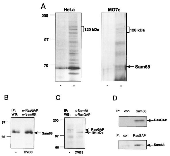
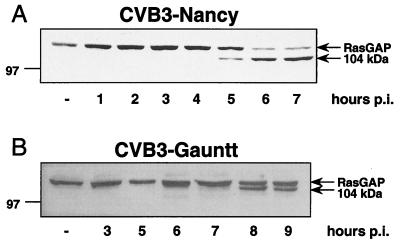
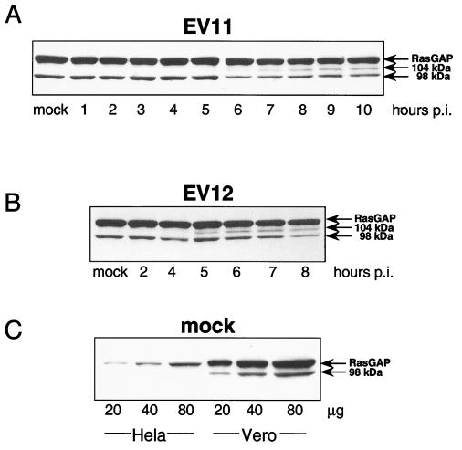
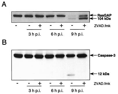
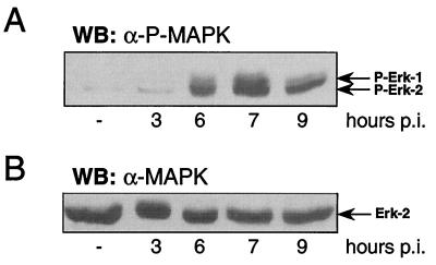
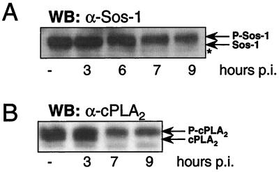
Recently, we reported on tyrosine phosphorylation of distinct cellular proteins in the course of enterovirus infections (M. Huber, H.-C. Selinka, and R. Kandolf, J. Virol. 71:595-600, 1997). These phosphorylation events were mediated by Src-like kinases and were shown to be necessary for effective virus replication. That study is now extended by examination of the interaction of the adapter protein Sam68, a cellular target of Src-like kinases which has been shown to interact with the poliovirus 3D polypeptide, with cellular signaling proteins as well as the function of the latter during infection. Here, we report that the RNA-binding and protein-binding protein Sam68 associates with the p21(ras) GTPase-activating protein RasGAP. Remarkably, RasGAP is cleaved during infections with different strains of coxsackievirus B3 as well as with echovirus 11 and echovirus 12, yielding a 104-kDa protein fragment. This cleavage event, which cannot be prevented by the general caspase inhibitor benzyloxycarbonyl-Val-Ala-Asp-fluoromethylketone, may promote the activation of the Ras pathway, as shown by the activating dual phosphorylation of the mitogen-activated protein kinases Erk-1 and Erk-2 in the late phase of infection. Moreover, downstream targets of the mitogen-activated protein kinases, i.e., the p21(ras) exchange factor Sos-1 and cytoplasmic phospholipase A2, are phosphorylated with parallel time courses during infection. Activation or inhibition of cellular signaling pathways may play a general role in regulating effective enterovirus replication and pathogenesis, and the results of this study begin to unravel the molecular cross talk between enterovirus infection and key cellular signaling networks.


Figures

FIG. 1
FIG. 1
Association of Sam68 and RasGAP in HeLa cells. (A) Multiple proteins associate with Sam68 in cells of human origin. Postnuclear supernatants of HeLa and MO7e cells were precleared and subsequently immunoprecipitated with anti-Sam68 antibodies (+). The precipitating agent (anti-mouse IgG antibodies coupled to agarose) served as a control (−). The precipitates were separated by SDS-PAGE and subjected to a silver staining procedure. Coprecipitated proteins in the range of 120 kDa are indicated. (B) Coimmunoprecipitation of Sam68 with antibodies specific for RasGAP. CVB3-infected (CVB3) or mock-infected (−) HeLa cells were lysed 5 h p.i., and postnuclear supernatants were immunoprecipitated (IP) with RasGAP-specific antibodies. The immunoprecipitates were analyzed on Western blots (WB) with Sam68-specific antibodies. Protein Sam68 is indicated. (C) RasGAP is coprecipitated with Sam68-specific antibodies. Postnuclear supernatants were immunoprecipitated with Sam68-specific antibodies and examined by Western blotting with RasGAP-specific antibodies. RasGAP and the 104-kDa cleavage product are indicated. (D) HeLa cell lysates were subjected to immunoprecipitation with anti-Sam68 antibodies or precipitating reagent (con) and with anti-RasGAP antibodies or precipitating reagent (con) and probed for RasGAP and Sam68, respectively. Sam68 and RasGAP are indicated.
FIG. 2
FIG. 2
Expression of RasGAP during the course of CVB3 infection. (A) RasGAP cleavage during the course of infection with CVB3 (Nancy strain). HeLa cells were infected with CVB3 (Nancy strain) or mock infected and lysed at 60-min intervals from 1 h to 7 h p.i. Protein preparations were subjected to Western blotting with RasGAP-specific antibodies. (B) RasGAP cleavage during the course of infection with CVB3 (Gauntt strain). HeLa cells were infected with CVB3 or mock infected and lysed from 3 h to 9 h p.i. Postnuclear supernatants were subjected to Western blotting with RasGAP-specific antibodies. RasGAP (120 kDa) and the 104-kDa protein are indicated.
FIG. 3
FIG. 3
RasGAP expression in EV11- and EV12-infected Vero cells. (A) Time course of expression of RasGAP in EV11-infected Vero cells. Vero cells were infected with EV11 and lysed at 60-min intervals from 1 h to 10 h p.i. Postnuclear supernatants were analyzed for RasGAP expression by Western blotting with RasGAP-specific antibodies. RasGAP, the 104-kDa protein, and the additional Vero cell-specific 98-kDa protein are indicated. (B) Time course of expression of RasGAP in EV12-infected Vero cells. Protein preparations of EV12- and mock-infected Vero cells were subjected to Western blotting with RasGAP-specific antibodies. RasGAP and RasGAP-related proteins of 104 and 98 kDa are indicated. (C) Comparison of RasGAP expression in noninfected HeLa and Vero cells. Lysates from HeLa and Vero cells (20, 40, and 80 μg) were separated by SDS–10% PAGE and probed with RasGAP-specific antibodies. RasGAP and the Vero cell-specific, RasGAP-related 98-kDa protein are indicated.
FIG. 4
FIG. 4
CVB3-induced RasGAP cleavage in the presence of the general caspase inhibitor ZVAD.fmk. (A) Effect of the caspase inhibitor ZVAD.fmk on RasGAP cleavage. HeLa cells were mock infected (leftmost lane) or infected with CVB3 for 3, 6, and 9 h in the presence (+) or absence (−) of 100 μM ZVAD.fmk. The cells were lysed, and postnuclear lysates were separated by SDS-PAGE. After transfer to a PVDF membrane, RasGAP was detected by Western blotting. RasGAP and the 104-kDa cleavage product are indicated. (B) Caspase-3 processing is prevented by ZVAD.fmk. The postnuclear lysates described for panel A were examined for caspase-3 expression by Western blotting. Caspase-3 is indicated.
FIG. 5
FIG. 5
Activation of MAPK during CVB3 infection. (A) MAPK are dually phosphorylated in CVB3-infected cells. HeLa cells were CVB3 (Gauntt strain) infected or mock infected (leftmost lane) and lysed at various times p.i. Postnuclear supernatants were separated by SDS-PAGE and subjected to Western blot (WB) analysis with antibodies specific for the dually phosphorylated MAPK Erk-1 and Erk-2. Phosphorylated Erk-1 and phosphorylated Erk-2 (P-Erk-1 and P-Erk-2, respectively) are indicated. (B) MAPK expression during CVB3 replication. The postnuclear lysates described for panel A were subjected to anti-MAPK Western blot analysis to confirm the presence of equal amounts of MAPK in these preparations. Erk-2 is indicated.
FIG. 6
FIG. 6
CVB3 replication induces the phosphorylation of MAPK substrates. (A) Hyperphosphorylation of Sos-1 during CVB3 infection. CVB3 (Gauntt strain)-infected and mock-infected (leftmost lane) HeLa cells were lysed at various times p.i., and postnuclear lysates were separated by SDS–8% PAGE and subjected to anti–Sos-1 Western blotting (WB). Sos-1 and phosphorylated Sos-1 (P-Sos-1) are indicated. Note the increasing distance between the Sos-1 protein and a cross-reacting protein (asterisk), which does not show a change in its electrophoretic mobility. (B) Mobility shift of cPLA2 during the course of CVB3 replication. The lysates described for panel A were analyzed by Western blotting with antibodies specific for cPLA2. cPLA2 and phosphorylated cPLA2 (P-cPLA2) are indicated.

References

    1. Andino R, Rieckhof G E, Achacoso P L, Baltimore D. Poliovirus RNA synthesis utilizes an RNP complex formed around the 5′-end of viral RNA. EMBO J. 1993;12:3587–3598. - PMC - PubMed
    1. Barbacid M. Ras genes. Annu Rev Biochem. 1987;56:779–827. - PubMed
    1. Bergelson J M, Cunningham J A, Droguett G, Kurt-Jones E A, Krithivas A, Hong J S, Horwitz M S, Crowell R L, Finberg R W. Isolation of a common receptor for coxsackie B viruses and adenoviruses 2 and 5. Science. 1997;275:1320–1323. - PubMed
    1. Bienz K, Egger D, Troxler M, Pasamontes L. Structural organization of poliovirus RNA replication is mediated by viral proteins of the P2 genomic region. J Virol. 1990;64:1156–1163. - PMC - PubMed
    1. Black T L, Safer B, Hovannessian A, Katze M G. The cellular 68,000-Mr protein kinase is highly autophosphorylated and activated yet significantly degraded during poliovirus infection: implications for translational regulation. J Virol. 1989;63:2244–2251. - PMC - PubMed

Publication types

MeSH terms