. 1999 May;73(5):3800-9.
doi: 10.1128/JVI.73.5.3800-3809.1999.

Expression and characterization of a novel structural protein of human cytomegalovirus, pUL25

Affiliations

Expression and characterization of a novel structural protein of human cytomegalovirus, pUL25

M C Battista et al. J Virol. 1999 May.

Abstract

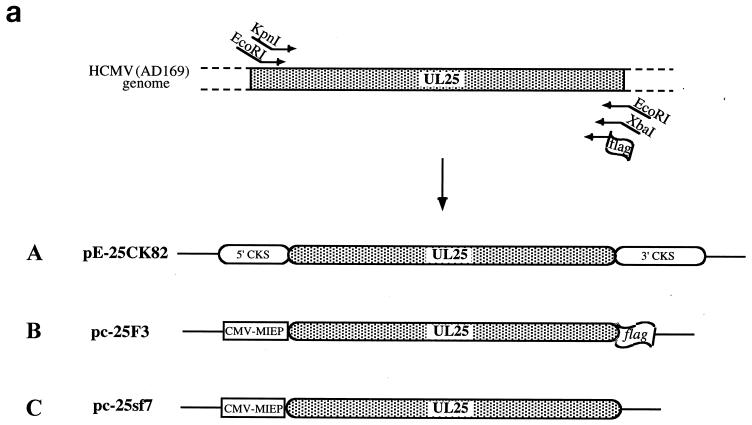
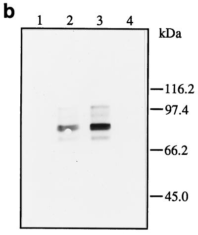
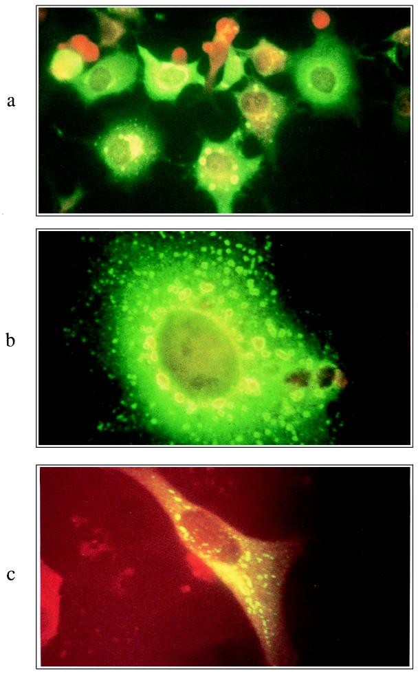
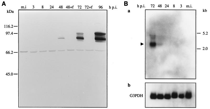
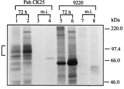
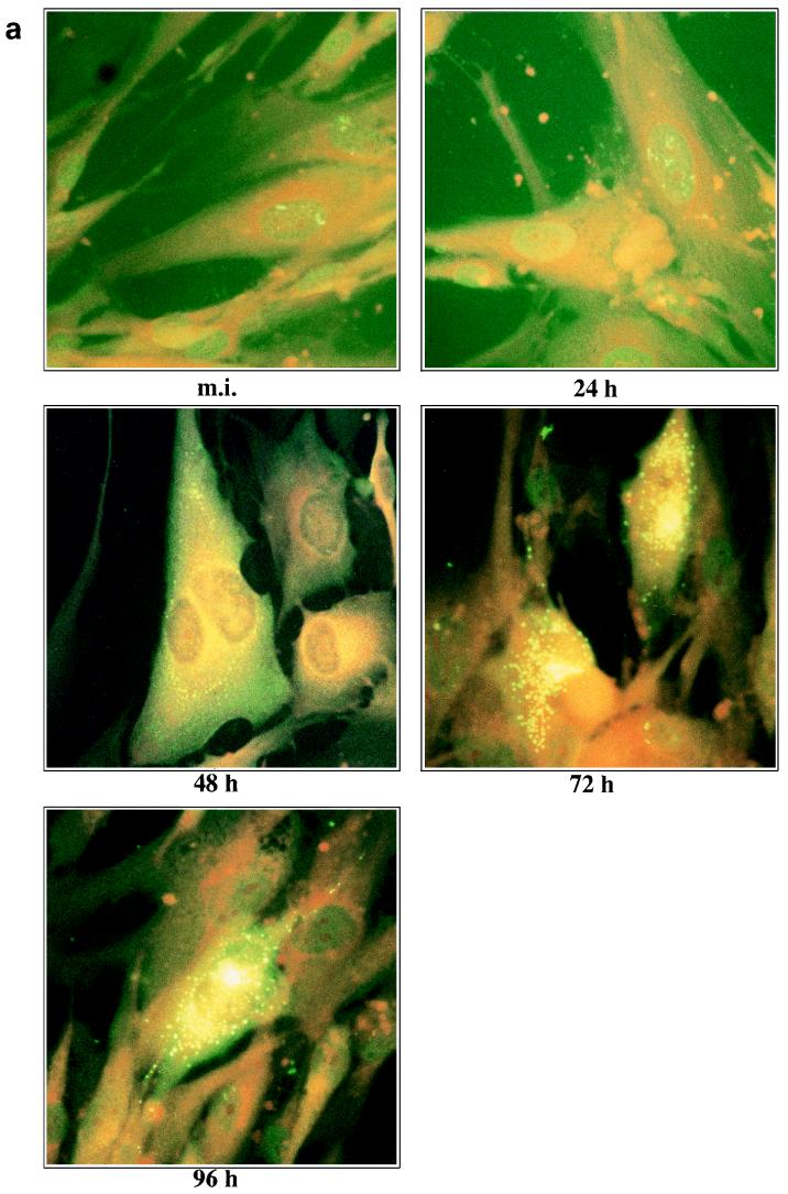
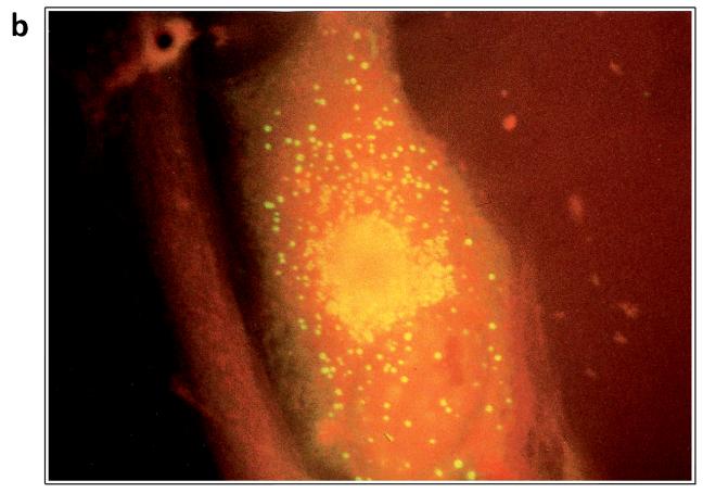
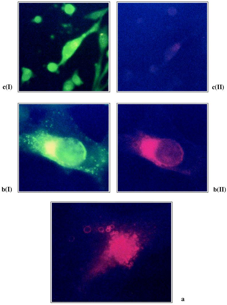
Human cytomegalovirus (HCMV) UL25 has recently been found to encode a new structural protein that is present in both virion and defective viral particles (C. J. Baldick and T. Shenk, J. Virol. 70:6097-6105, 1996). In the present work a polyclonal antibody was raised against a prokaryotic pUL25 fusion protein in order to investigate the biosynthesis and localization of the UL25 product (pUL25) during HCMV replication in human fibroblasts. Furthermore, pUL25 was transiently expressed in its native form and fused to the FLAG epitope, in COS7 and U373MG cells, in order to compare the properties of the isolated protein and that produced during infection. Immunoblotting analysis revealed a group of polypeptides, ranging from 80 to 100 kDa, in both transfected and infected cells; in vivo labeling experiments with infected cells demonstrated they are posttranslationally modified by phosphorylation. The transcriptional analysis of the UL25 open reading frame combined with the study of pUL25 biosynthesis showed true late kinetics for this protein in infected human fibroblasts. By indirect immunofluorescence both recombinant and viral pUL25 were detected exclusively in the cytoplasm of transfected or infected cells. Interestingly, pUL25 was shown to localize in typical condensed structures in the perinuclear region as already observed for other HCMV tegument proteins. Colocalization of ppUL99 in the same vacuoles suggests that these structure are endosomal cisternae, which are proposed to be a preferential site of viral particle envelopment. Our data suggest that pUL25 is most likely a novel tegument protein and possibly plays a key role in the process of envelopment.


Figures

FIG. 1
FIG. 1
(a) Construction of UL25 expression plasmids. The different pairs of primers used for the amplification of ORF UL25 from the AD169 strain genome are shown at the top. A, prokaryotic expression plasmid; B, pUL25-FLAG eukaryotic expression plasmid; C, native pUL25 eukaryotic expression plasmid. MIEP, major immediate-early promoter. (b) IB analysis of pUL25-FLAG transiently expressed in mammalian cells (48 h posttransfection). Proteins of whole-cell lysates were separated by SDS–9% PAGE, transferred to nitrocellulose, and probed with the anti-FLAG monoclonal antibody M2 (diluted 1:200). Lanes 1 and 2, U373MG cells transiently transfected with control vector pcDNA3 and pc-25F3, respectively; lanes 3 and 4, COS7 cells transiently transfected with pc-25F3 and pcDNA3, respectively. Positions of molecular mass markers are shown at the right.
FIG. 1
FIG. 1
(a) Construction of UL25 expression plasmids. The different pairs of primers used for the amplification of ORF UL25 from the AD169 strain genome are shown at the top. A, prokaryotic expression plasmid; B, pUL25-FLAG eukaryotic expression plasmid; C, native pUL25 eukaryotic expression plasmid. MIEP, major immediate-early promoter. (b) IB analysis of pUL25-FLAG transiently expressed in mammalian cells (48 h posttransfection). Proteins of whole-cell lysates were separated by SDS–9% PAGE, transferred to nitrocellulose, and probed with the anti-FLAG monoclonal antibody M2 (diluted 1:200). Lanes 1 and 2, U373MG cells transiently transfected with control vector pcDNA3 and pc-25F3, respectively; lanes 3 and 4, COS7 cells transiently transfected with pc-25F3 and pcDNA3, respectively. Positions of molecular mass markers are shown at the right.
FIG. 2
FIG. 2
IIF analysis of pUL25-FLAG transiently expressed in mammalian cells (48 h posttransfection), using the anti-FLAG monoclonal antibody M2 (diluted 1:200). (a and b) COS7/pc-25F3; (c) U373MG/pc-25F3. Magnification, ×40 (a) and ×100 (b and c).
FIG. 3
FIG. 3
(A) Time course of pUL25 expression during the HCMV replication cycle, determined by IB analysis. Whole-cell lysates of mock-infected (m.i.) and HCMV-infected HEL cells at various times after infection were separated by SDS–9% PAGE, and the proteins were then transferred to nitrocellulose and immunostained with the mouse ascites fluid PAb CK25 (diluted 1:500). +f, samples treated with foscarnet (100 μg/ml) after adsorption of virus. Positions of molecular mass markers are shown on the left. (B) Northern blot analysis of the UL25 transcript. Total RNA was prepared from mock-infected (m.i.) and HCMV-infected HEL cells at various times after infection by using an RNAzol B kit. Aliquots of 15 μg of total RNA were subjected to electrophoresis through a 1% agarose gel containing 2.2 M formaldehyde and then transferred to nylon. The resulting filter was hybridized with a 32P-labeled probe of the entire UL25 sequence (a) and rehybridized with a 32P-labeled G3PDH gene probe (b). The location of the 2.4-kb UL25 transcript is indicated by the arrowhead. The molecular sizes of 28S and 18S human rRNAs are indicated on the right.
FIG. 4
FIG. 4
In vivo phosphorylation of mock-infected (m.i.) and HCMV-infected human fibroblasts. Labeling was performed with either 33Pi (lanes 1, 3, 5, and 7) or [35S]cysteine (lanes 2, 4, 6, and 8) from 48 to 72 h after infection with HCMV strain AD169. Cell lysates were immunoprecipitated with either the mouse ascites fluid PAb CK25 or anti-pUL83 (pp65) monoclonal antibody 9220. The bracket on the left shows the group of polypeptides specifically detected by PAb CK25. Positions of molecular mass markers are shown on the right.
FIG. 5
FIG. 5
(a) Subcellular localization of pUL25 during the HCMV replication cycle by IIF analysis of mock-infected (m.i.) and HCMV-infected HEL cells, using the mouse ascites fluid PAb CK25 (diluted 1:400). Magnification, ×40. (b) Association of pUL25 with cap-like structures. IIF analysis of HCMV-infected human fibroblasts (96 h p.i.) with PAb CK25 is shown. Magnification, ×100.
FIG. 5
FIG. 5
(a) Subcellular localization of pUL25 during the HCMV replication cycle by IIF analysis of mock-infected (m.i.) and HCMV-infected HEL cells, using the mouse ascites fluid PAb CK25 (diluted 1:400). Magnification, ×40. (b) Association of pUL25 with cap-like structures. IIF analysis of HCMV-infected human fibroblasts (96 h p.i.) with PAb CK25 is shown. Magnification, ×100.
FIG. 6
FIG. 6
(a) Subcellular localization of ppUL99 in human fibroblasts at 104 h after HCMV infection. Cells were incubated with the monoclonal antibody P2G11 and than with the secondary TRITC-conjugated antibody. (b) Subcellular localization of ppUL25 in HEL at 104 h p.i. I, an optical filter allowing the view of the secondary fluorescein-conjugated antibody, which specifically recognizes the ascites fluid PAb CK25, was used. II, subcellular localization of ppUL99 in the same cell, with an optical filter allowing the view of the TRITC-conjugated antibody, which specifically recognizes the monoclonal P2G11. Green and red staining coincide, thus showing that in this phase of the infection cycle, ppUL25 and ppUL99 colocalize in the same subcellular compartment. (c) Cells incubated as for panel b except for the omission of the monoclonal antibody P2G11. Thus, the green filter showed the localization of ppUL25 (I), while the red filter showed that no red staining was present (II), proving that the TRITC-conjugated antibody recognized exclusively the monoclonal P2G11.

References

    1. Baldick C J, Jr, Shenk T. Proteins associated with purified human cytomegalovirus particles. J Virol. 1996;70:6097–6105. - PMC - PubMed
    1. Battista M C, Bergamini G, Campanini F, Landini M P, Ripalti A. Intracellular production of a major cytomegalovirus antigenic protein in the methylotrophic yeast Pichia pastoris. Gene. 1996;176:197–201. - PubMed
    1. Bogner E, Reschke M, Reis B, Mockenhaupt T, Radsak K. Identification of the gene product encoded by ORF UL56 of the human cytomegalovirus genome. Virology. 1993;196:290–293. - PubMed
    1. Bolling T J, Mandecki M. An Escherichia coli expression vector for high-level production of heterologous proteins in fusion with CMP-KDO synthetase. BioTechniques. 1990;8:488–490. - PubMed
    1. Bradshaw P A, Duran-Guarino M R, Perkins S, Rowe J I, Fernandez J, Fry K E, Reyes G R, Young L, Foung S K. Localization of antigenic sites on human cytomegalovirus virion structural proteins encoded by UL48 and UL56. Virology. 1994;205:321–328. - PubMed

Publication types

LinkOut - more resources