. 1999 May;73(5):3994-4003.
doi: 10.1128/JVI.73.5.3994-4003.1999.

Gene therapy vectors based on adeno-associated virus type 1

Affiliations

Gene therapy vectors based on adeno-associated virus type 1

W Xiao et al. J Virol. 1999 May.

Abstract

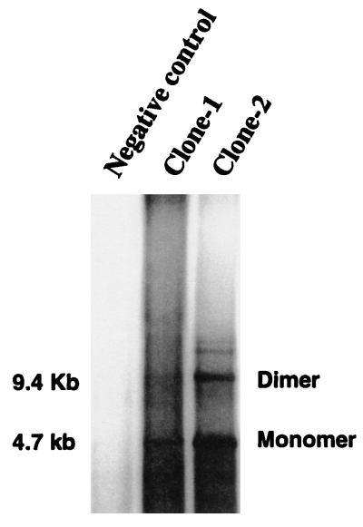
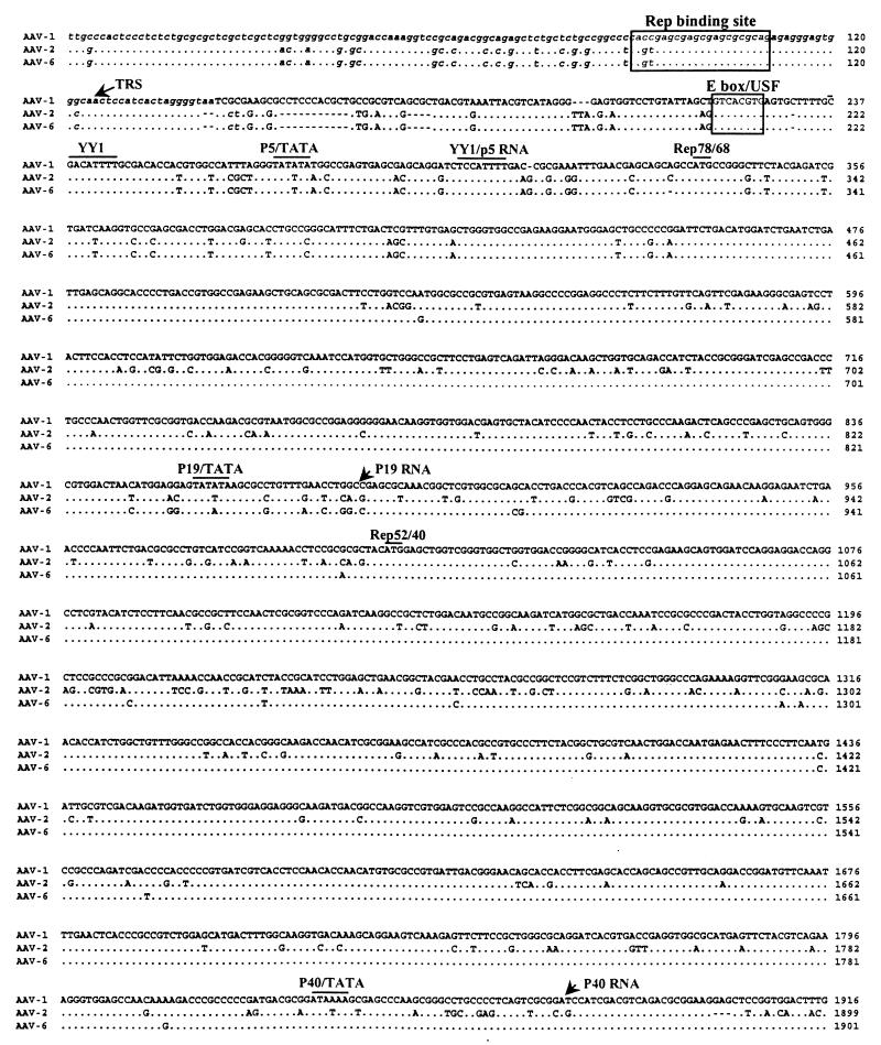
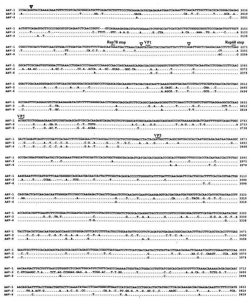
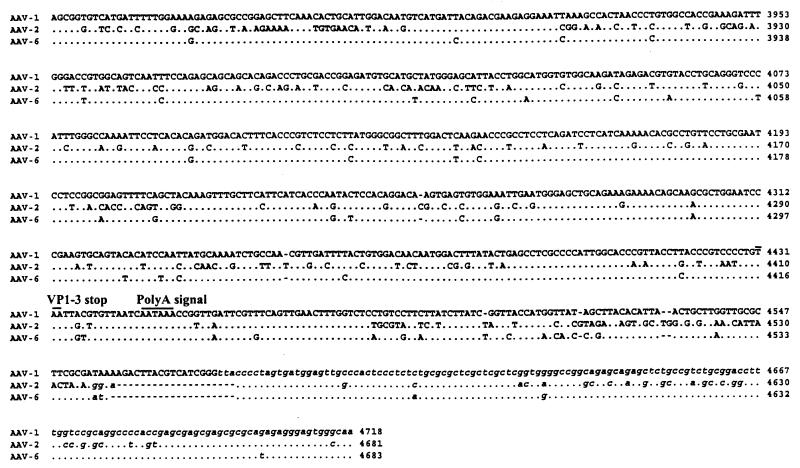
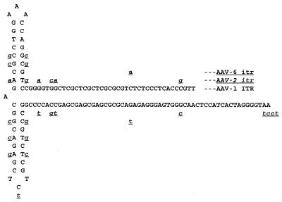
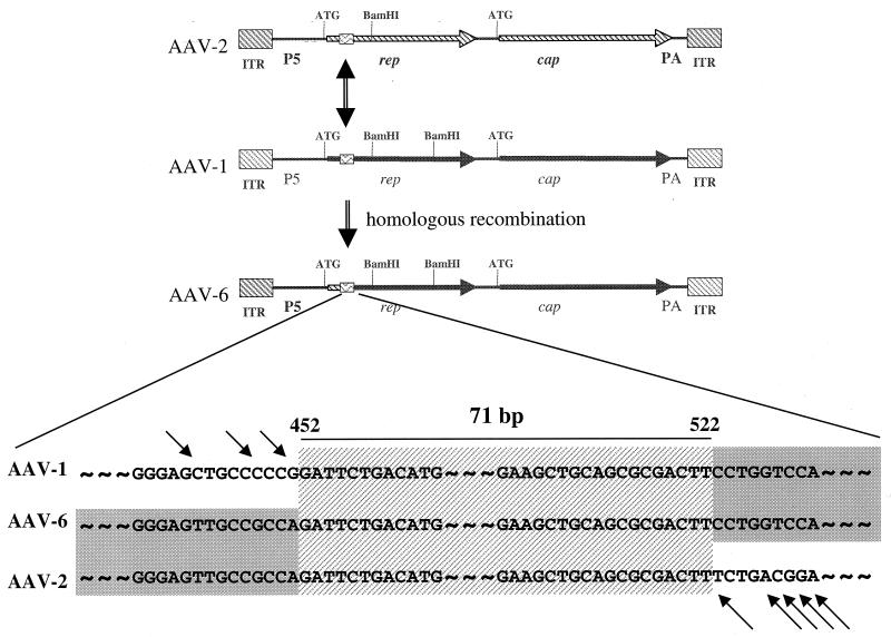
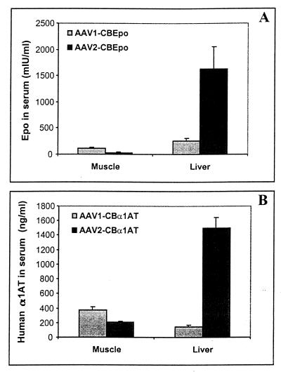
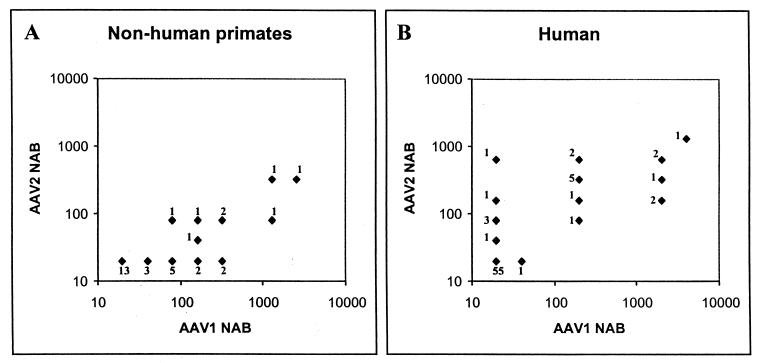
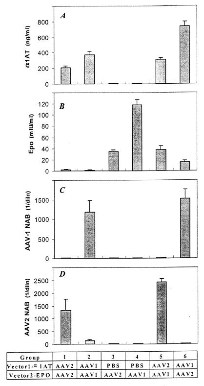
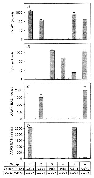
The complete sequence of adeno-associated virus type 1 (AAV-1) was defined. Its genome of 4,718 nucleotides demonstrates high homology with those of other AAV serotypes, including AAV-6, which appears to have arisen from homologous recombination between AAV-1 and AAV-2. Analysis of sera from nonhuman and human primates for neutralizing antibodies (NAB) against AAV-1 and AAV-2 revealed the following. (i) NAB to AAV-1 are more common than NAB to AAV-2 in nonhuman primates, while the reverse is true in humans; and (ii) sera from 36% of nonhuman primates neutralized AAV-1 but not AAV-2, while sera from 8% of humans neutralized AAV-2 but not AAV-1. An infectious clone of AAV-1 was isolated from a replicated monomer form, and vectors were created with AAV-2 inverted terminal repeats and AAV-1 Rep and Cap functions. Both AAV-1- and AAV-2-based vectors transduced murine liver and muscle in vivo; AAV-1 was more efficient for muscle, while AAV-2 transduced liver more efficiently. Strong NAB responses were detected for each vector administered to murine skeletal muscle; these responses prevented readministration of the same serotype but did not substantially cross-neutralize the other serotype. Similar results were observed in the context of liver-directed gene transfer, except for a significant, but incomplete, neutralization of AAV-1 from a previous treatment with AAV-2. Vectors based on AAV-1 may be preferred in some applications of human gene therapy.


Figures

FIG. 1
FIG. 1
DNA hybridization analysis of the rescue and replication of the AAV-1 infectious clone. Two independent clones of pAAV-1 (Clone-1 and Clone-2) were transfected into 293 cells, which were then superinfected with adenovirus type 5. Hirt DNA was extracted 48 h postinfection and digested with DpnI before electrophoresis in an 0.8% agarose gel. An internal BamHI fragment of pAAV-1 was used as a probe. The autoradiograph shows the locations of replicated dimers and monomers and their molecular sizes. The analysis included cells not infected (negative control) or infected with clone-1 or clone-2.
FIG. 2
FIG. 2
Alignment of nucleotides for AAV-1, AAV-2, and AAV-6. The full sequence of AAV-1 is indicated in the top line; the AAV-2 and AAV-6 sequences are shown below with nucleotide differences. Homology is indicated by dots. Critical landmarks in the structures of the AAV genomes are shown. Gaps are indicated by dashes. The 3′ ITR of AAV-1 is shown in the same configuration as in the published AAV-2 and AAV-6 sequences (22). TRS, terminal resolution site.
FIG. 2
FIG. 2
Alignment of nucleotides for AAV-1, AAV-2, and AAV-6. The full sequence of AAV-1 is indicated in the top line; the AAV-2 and AAV-6 sequences are shown below with nucleotide differences. Homology is indicated by dots. Critical landmarks in the structures of the AAV genomes are shown. Gaps are indicated by dashes. The 3′ ITR of AAV-1 is shown in the same configuration as in the published AAV-2 and AAV-6 sequences (22). TRS, terminal resolution site.
FIG. 3
FIG. 3
Potential secondary structure of AAV-1 ITR. Nucleotide differences in the corresponding sequences of AAV-2 and AAV-6 relative to AAV-1 are underlined. AAV-2 sequence differences are lowercase, underlined, and italicized, whereas AAV-6 sequence differences are lowercase and underlined.
FIG. 4
FIG. 4
Hypothesis for the creation of AAV-6 from homologous recombination between AAV-1 and AAV-2. The first line represents the AAV-2 genome, with Rep and Cap sequences demarcated by hatched arrows. The second line represents the AAV-1 genome, with the Rep and Cap sequences depicted as solid arrows. The hypothesis suggests homologous recombination between AAV-2 and AAV-1 at the region of the box containing wavy lines; this region contains a highly homologous sequence of the Rep gene shared by AAV-1 and AAV-2. PA, poly(A). A more detailed illustration of the common region of these three viruses is shown at the bottom. Arrows indicate nucleotides that differ.
FIG. 5
FIG. 5
In vivo activities of AAV-1 and AAV-2 vectors. Recombinant stocks of virus were generated by transfection and purified through cesium chloride gradients. (A) AAV-1 and AAV-2 vectors expressing murine erythropoietin (Epo) from a cytomegalovirus-enhanced β-actin promoter (CB). Equivalent stocks of these two vectors were injected into muscle (5 × 10¹⁰ genomes) or liver via the portal circulation (1 × 10¹¹ genomes), and the animals (four groups) were analyzed on day 30 for the presence of erythropoietin in blood. Data for the groups were combined. Error bars show standard errors. (B) An identical experiment was performed with AAV-1 and AAV-2 vectors expressing human α1-antitrypsin (α1AT) from the same promoter. Serum harvested from four groups of animals 30 days after gene transfer was analyzed for the presence of α1-antitrypsin by an ELISA. Data are reported as in panel A.
FIG. 6
FIG. 6
NAB to AAV-1 and AAV-2 in nonhuman and human primates. Sera were analyzed for neutralizing activity against rAAV-1 and rAAV-2 as described in Materials and Methods. The reciprocal dilution of the NAB was plotted for AAV-2 versus AAV-1. Sera were titrated in sequential twofold dilutions. A titer of 1:20 represents the background (i.e., no detectable NAB). The number next to each datum point represents the total number of animals or humans with a particular NAB profile. In panel A, 33 adolescent rhesus monkeys were analyzed. In panel B, 77 normal human volunteers were evaluated.
FIG. 7
FIG. 7
Readministration of AAV vectors in muscle. C57BL/6 mice were evaluated for AAV-mediated gene transfer following introduction into naive animals as well as 30 days following the first vector administration. The different experimental groups are shown below the graphs. In each case, vector 1 expressed α1-antitrypsin (α1AT) from a cytomegalovirus-enhanced β-actin promoter, while vector 2 expressed murine erythropoietin (EPO) from the same promoter. Group 1, AAV-2 followed by AAV-2; group 2, AAV-1 followed by AAV-1; group 3, phosphate-buffered saline (PBS) followed by AAV-2; group 4, PBS followed by AAV-1; group 5, AAV-2 followed by AAV-1; group 6, AAV-1 followed by AAV-2. (A) Serum α1-antitrypsin 30 days after vector 1 administration. (B) Serum erythropoietin (Epo) measured by an ELISA 30 days after vector 2 administration. (C) Reciprocal dilution (diln) of NAB to AAV-1 at day 30. (D) Reciprocal dilution of NAB to AAV-2 at day 30.
FIG. 8
FIG. 8
Readministration of AAV vectors in the liver. These experiments are identical to those described in the legend to Fig. 7, except that animals received vectors in the portal circulation to target the liver.

References

    1. Balague C, Kalla M, Zhang W W. Adeno-associated virus Rep78 protein and terminal repeats enhance integration of DNA sequences into the cellular genome. J Virol. 1997;71:3299–3306. - PMC - PubMed
    1. Bantel-Schaal U, zur Hausen H. Characterization of the DNA of a defective human parvovirus isolated from a genital site. Virology. 1984;134:52–63. - PubMed
    1. Berns K I. Parvoviridae: the viruses and their replication. In: Fields B N, Knipe D M, Howley P M, editors. Fundamental virology. 3rd ed. Vol. 2. Philadelphia, Pa: Lippincott-Raven Publishers; 1995. pp. 1007–1041.
    1. Chiorini J A, Yang L, Liu Y, Safer B, Kotin R M. Cloning of adeno-associated virus type 4 (AAV4) and generation of recombinant AAV4 particles. J Virol. 1997;71:6823–6833. - PMC - PubMed
    1. Clark K R, Voulgaropoulou F, Fraley D M, Johnson P R. Cell lines for the production of recombinant adeno-associated virus. Hum Gene Ther. 1995;6:1329–1341. - PubMed

Publication types

MeSH terms

Associated data