. 1999 May;73(5):4052-61.
doi: 10.1128/JVI.73.5.4052-4061.1999.

In vitro infection of human peripheral blood mononuclear cells by GB virus C/Hepatitis G virus

Affiliations

In vitro infection of human peripheral blood mononuclear cells by GB virus C/Hepatitis G virus

M Fogeda et al. J Virol. 1999 May.

Abstract

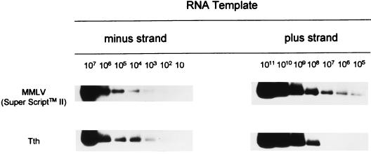
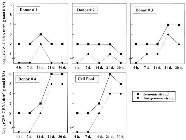
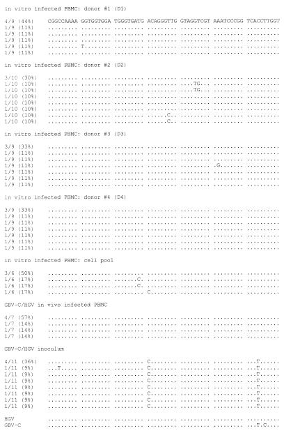
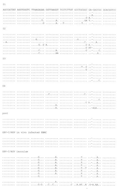
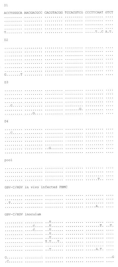
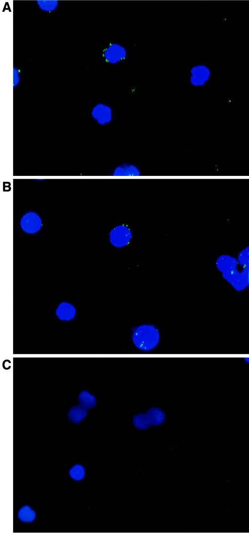
GB virus C (GBV-C), also known as hepatitis G virus, is a recently discovered flavivirus-like RNA agent with unclear pathogenic implications. To investigate whether human peripheral blood mononuclear cells (PBMC) are susceptible to in vitro GBV-C infection, we have incubated PBMC from four healthy blood donors with a human GBV-C RNA-positive serum. By means of (i) strand-specific reverse transcription-PCR, cloning, and sequencing; (ii) sucrose ultracentrifugation and RNase sensitivity assays; (iii) fluorescent in situ hybridization; and (iv) Western blot analysis, it has been demonstrated that GBV-C is able to infect in vitro cells and replicate for as long as 30 days under the conditions developed in our cell culture system. The concentration of GBV-C RNA increased during the second and third weeks of culture. The titers of the genomic strand were 10 times higher than the titers of the antigenomic strand. In addition, the same predominant GBV-C sequence was found in all PBMC cultures and in the in vivo-GBV-C-infected PBMC isolated from the donor of the inoculum. GBV-C-specific fluorescent in situ hybridization signals were confined to the cytoplasm of cells at different times during the culture period. Finally, evidence obtained by sucrose ultracentrifugation, RNase sensitivity assays, and Western blot analysis of the culture supernatants suggests that viral particles are released from in vitro-GBV-C-infected PBMC. In conclusion, our study has demonstrated, for the first time, GBV-C replication in human lymphoid cells under experimental in vitro infection conditions.


Figures

FIG. 1
FIG. 1
Sensitivity and specificity of the RT-PCR assay for the detection of genomic and antigenomic GBV-C RNA strands. Synthetic GBV-C RNA transcripts (corresponding to the 5′ NC region) of positive and negative polarity were generated by in vitro transcription, and 10-fold dilutions were performed in polyethylene glycol- and diethylpyrocarbonate-treated water. cDNA synthesis was performed in the presence of the sense primer, and afterward the reverse transcriptase was inactivated by heating the product at 95°C for 45 min (Moloney murine leukemia virus [MMLV] Super Script II) or chelation (Tth). The number of target template copies was determined from the optical density measurement and confirmed by electrophoresis in an agarose gel. Assays included amplification of 0 to 106 RNA copies per reaction. Subsequently, the products of the nested PCRs were analyzed by Southern hybridization with a 32P-labelled probe.
FIG. 2
FIG. 2
Determination of GBV-C RNA content in experimentally GBV-C-infected PBMC. Results for each individual healthy blood donor and the cell pool are expressed as the log10 of the genomic and antigenomic GBV-C RNA contents per microgram of total RNA from cells, as determined with successive 10-fold dilutions at the end of the 4-h infection period and after 7, 14, 21, and 30 days of culture.
FIG. 3
FIG. 3
Alignment of the GBV-C/HGV 5′ NC sequences (255 bp; nt −632 to −378, according to R10291 isolate numbering [18]) amplified from in vitro-infected PBMC (D1, D2, D3, D4, and cell pool), in vivo-infected PBMC isolated from the patient whose serum was used as the inoculum, and the GBV-C/HGV-positive serum used as the inoculum. Sequence identity with the predominant sequence found in the in vitro-infected cells is indicated by dots, and insertions are indicated by dashes. Comparisons with HGV (R10291 [18]) and GBV-C (U36380 [17]) prototypes are shown. The pair of numbers on the left of each line, one before and one after the shill, indicates the number of clones analyzed and the percentages of each sequence within the spectrum obtained, respectively.
FIG. 3
FIG. 3
Alignment of the GBV-C/HGV 5′ NC sequences (255 bp; nt −632 to −378, according to R10291 isolate numbering [18]) amplified from in vitro-infected PBMC (D1, D2, D3, D4, and cell pool), in vivo-infected PBMC isolated from the patient whose serum was used as the inoculum, and the GBV-C/HGV-positive serum used as the inoculum. Sequence identity with the predominant sequence found in the in vitro-infected cells is indicated by dots, and insertions are indicated by dashes. Comparisons with HGV (R10291 [18]) and GBV-C (U36380 [17]) prototypes are shown. The pair of numbers on the left of each line, one before and one after the shill, indicates the number of clones analyzed and the percentages of each sequence within the spectrum obtained, respectively.
FIG. 4
FIG. 4
FISH in experimentally GBV-C-infected cells maintained in culture for 30 days. The fluorescent signals were always located in the cytoplasm of the cells. Shown are positive signals obtained in cells from donor 4 at days 7 (A) and 30 (B) of culture, as well as the absence of a fluorescent signal in cells from a negative-control culture (cell pool inoculated with GBV-C-negative serum at day 30) (C). Cells were counterstained with propidium iodide. Original magnifications, ×650.
FIG. 5
FIG. 5
Western blot analysis of culture supernatants. Culture supernatants of cells from donor 4 at days 21 (lanes 1 to 3) and 30 (lanes 4 to 6), and 100 ng of a recombinant putative E2 protein of GBV-C (lane 7), were subjected to polyacrylamide gel electrophoresis and Western blot analysis, using as primary antibodies a MAb prepared against the E2 protein of GBV-C (27), at a final concentration of 1 μg/ml (A); a 1:200 dilution of a GBV-C RNA-negative human serum with detectable circulating antibodies to the putative E2 protein of GBV-C (B); and a 1:200 dilution of a GBV-C RNA-negative human serum without detectable antibodies to the E2 protein of GBV-C (C). Lanes: 1 and 4, pellets frozen immediately after ultracentrifugation; 2 and 5, pellets treated with 0.1% NP-40 for 3 h at room temperature and immediately frozen; 3 and 6, pellets treated with NP-40 and ultracentrifuged again.

References

    1. Aikawa T, Sugay Y, Okamoto H. Hepatitis G infection in drug abusers with chronic hepatitis C. N Engl J Med. 1996;334:195. - PubMed
    1. Alter H J, Nakatsuyi Y, Melpolder J, Wages J, Wesley R, Shih J W K, Kim J P. The incidence of transfusion-associated hepatitis G virus infection and its relation to liver disease. N Engl J Med. 1997;336:747–754. - PubMed
    1. Berenguer M, Terrault N A, Piatak M, Yun A, Kim J P, Lau J Y N, Lake J R, Roberts J R, Ascher N, Ferrell L, Wright T. Hepatitis G virus infection in patients with hepatitis C virus infection undergoing liver transplantation. Gastroenterology. 1996;111:1569–1575. - PubMed
    1. Bukh J, Kim J P, Govindarajan S, Apgar C L, Foung S K H, Wages J, Jr, Yun A J, Shapiro M, Emerson S U, Purcell R H. Experimental infection of chimpanzees with hepatitis G virus and genetic analysis of the virus. J Infect Dis. 1998;177:855–862. - PubMed
    1. Chomzynski P, Sacchi N. Single-step method of RNA isolation by acid guanidinium thiocyanate-phenol-chloroform extraction. Anal Biochem. 1987;162:156–159. - PubMed

Publication types

Associated data

LinkOut - more resources