. 1999 May;73(5):4074-82.
doi: 10.1128/JVI.73.5.4074-4082.1999.

Mutational analysis of glycosylation, membrane translocation, and cell surface expression of the hepatitis E virus ORF2 protein

Affiliations

Mutational analysis of glycosylation, membrane translocation, and cell surface expression of the hepatitis E virus ORF2 protein

M Zafrullah et al. J Virol. 1999 May.

Abstract

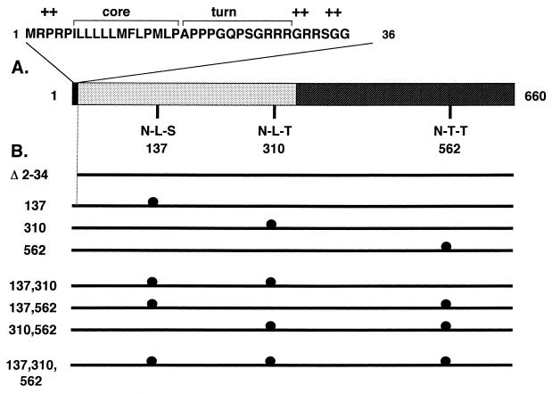
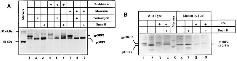
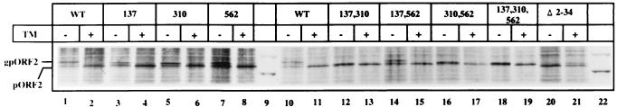
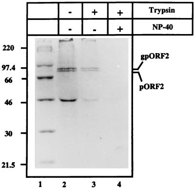
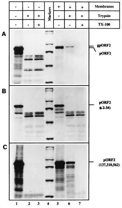
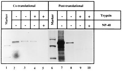
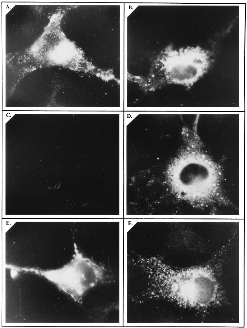
Hepatitis E virus (HEV) is the etiological agent for viral hepatitis type E, which is a major problem in the developing world. Because HEV cannot be cultured in vitro, very little information exists on the mechanisms of HEV gene expression and genome replication. HEV is a positive-strand RNA virus with three potential open reading frames (ORFs), one of which (ORF2) is postulated to encode the major viral capsid protein (pORF2). We earlier showed (S. Jameel, M. Zafrullah, M. H. Ozdener, and S. K. Panda, J. Virol. 70:207-216, 1996) pORF2 to be a approximately 88-kDa glycoprotein, carrying N-linked glycans and a potential endoplasmic reticulum (ER)-directing signal at its N terminus. Treatment with the drugs brefeldin A and monensin suggest that the protein may accumulate within the ER. Based on mutational analysis, we demonstrate Asn-310 to be the major site of N-glycan addition. In COS-1 cell expression and in vitro translation experiments, we confirm the ER-translocating nature of the pORF2 N-terminal hydrophobic sequence and show that the protein is cotranslationally, but not posttranslationally, translocated across the ER membrane. Earlier, we had also demonstrated cell surface localization of a fraction of the COS-1 cell-expressed pORF2. Using glycosylation- and translocation-defective mutants of pORF2, we now show that while transit of pORF2 into the ER is necessary for its cell surface expression, glycosylation of the protein is not required for such localization. These results may offer clues to the mechanisms of gene expression and capsid assembly in HEV.


Figures

FIG. 1
FIG. 1
ORF2 features and mutants. (A) The 660-amino-acid protein (pORF2) encoded by HEV ORF2 is shown schematically, with two major regions: the extreme N terminus, containing the putative signal sequence (filled), and the basic highly N-terminal half of the protein, containing about 10% arginine residues (dotted). The extreme N-terminal region is expanded to show three domains characteristic of eukaryotic signal sequences, the hydrophobic core domain, a region containing turn-inducing amino acids, and a positively charged domain (+). The positions of three potential N-linked glycosylation sites (N-X-S/T) are shown at amino acid residues 137, 310, and 562 in the primary sequence of pORF2. (B) Various mutants used in this study are shown schematically. The Δ2-34 mutant carries a 5′-end deletion in ORF2 corresponding to amino acids 2 to 34 in the polypeptide sequence. The glycosylation mutants with Asn→Ala changes at the indicated positions are shown (●). The nomenclature corresponds to the amino acid position(s) changed in that particular mutant.
FIG. 2
FIG. 2
Effects of brefeldin A and monensin on pORF2 expression. (A) COS-1 cells transfected with pSG-ORF2 were pulse-labeled with [35S]promix for 30 min, followed by a 3-h chase in complete DMEM. The cells were left untreated (lanes 1 and 3) or treated with 10 μg of brefeldin A (lanes 4 to 6)/ml, 5 μM monensin (lanes 7 to 9), or 10 μg of tunicamycin (lanes 2, 5, and 8)/ml during the prelabeling, pulse-labeling, and chase periods. Cell lysates were prepared, and the polypeptides were immunoprecipitated with a rabbit anti-pORF2 antiserum. Washed immunoprecipitates were subjected to mock treatment (lanes 1, 2, 4, 5, 7, and 8) or Endo H digestion (lanes 3, 6, and 9), and the polypeptides were analyzed on an SDS–7.5% polyacrylamide gel, followed by fluorography. Various treatments (+), the positions of gpORF2 and pORF2, and molecular size markers are indicated. (B) COS-1 cells transfected with pSG-ORF2 (lanes 1 to 4) or pSG-ORF2[Δ2-34] (lanes 6 to 9) were labeled with [35S]promix for 4 h in the absence (lanes 1, 2, 6, and 7) or presence (lanes 3, 4, 8, and 9) of 2.5 μg of brefeldin/ml. Following immunoprecipitation, the polypeptides in the precipitates were mock treated (lanes 1, 3, 5, and 7) or digested with Endo H (lanes 2, 4, 6, and 8) and analyzed on an SDS–7.5% polyacrylamide gel, followed by fluorography. The positions of gpORF2 and pORF2 are indicated. Only molecular size markers corresponding to 97.4 and 66 kDa are shown.
FIG. 3
FIG. 3
Expression and glycosylation of mutant pORF2 proteins. COS-1 cells transfected with pSG-ORF2 (lanes 1, 2, 10, and 11), pSG-ORF2[Asn→Ala mutants] (lanes 3 to 8 and 12 to 19), or pSG-ORF2[Δ2-34] (lanes 20 and 21) were labeled with [35S]promix for 4 h. Tunicamycin (10 μg/ml) was absent (−) or present (+) during the prelabeling and labeling periods as indicated. After immunoprecipitation, the polypeptides were analyzed on a SDS–7.5% polyacrylamide gel, followed by fluorography. The positions of gpORF2 and pORF2 are indicated. Only molecular size markers corresponding to 97.4 and 66 kDa are shown (lanes 9 and 22). WT, wild type; TM, tunicamycin.
FIG. 4
FIG. 4
Microsomal localization of pORF2. COS-1 cells transfected with pSG-ORF2 were labeled with [35S]promix for 4 h and used to prepare the microsomal fraction as described in Materials and Methods. The microsomal fraction was then incubated in the absence (−) or presence (+) of 25 μg of trypsin/ml and/or 0.5% NP-40, on ice for 60 min. Following immunoprecipitation, the polypeptides were analyzed on an SDS–10% polyacrylamide gel, followed by fluorography. The positions of gpORF2 and pORF2 are indicated. Molecular size markers are shown (lane 1).
FIG. 5
FIG. 5
In vitro synthesis and translocation of pORF2 and its mutants. Coupled transcription-translation reactions were carried out by the TNT T7 system (Promega) programmed with plasmid pSG-ORF2 (A), pSG-ORF2[Δ2-34] (B), or pSG-ORF2[137,310,562] (C). Canine pancreatic membranes were absent (lanes 1 to 3) or present (lanes 4 to 7) during the in vitro reaction. Subsequently, each reaction mix was divided into three parts and either mock treated (lanes 1 and 5) or treated directly with 250 μg of trypsin/ml in the absence (lanes 2 and 6) or presence (lanes 3 and 7) of 0.7% Triton X-100. The protected polypeptides were analyzed on an SDS–7.5% polyacrylamide gel, followed by fluorography. Molecular size markers (lane 4) are shown as in Fig. 4. The positions of gpORF2, pORF2, pORF2[Δ2-34], and pORF2[137,310,562] are indicated.
FIG. 6
FIG. 6
Co- and posttranslational translocation of pORF2. Plasmid pSG-ORF2 was subjected to in vitro-coupled transcription-translation. Following co- or posttranslational translocation of the in vitro-synthesized protein, as described in Materials and Methods, the membranes were pelleted down through a 250 mM sucrose–TBS cushion, resuspended in TBS, and divided into three equal aliquots. The aliquots were either mock treated (lanes 3 and 8) or treated with 25 μg trypsin/ml in the absence (lanes 4 and 9) or presence (lanes 5 and 10) of 0.5% NP-40. Input reactions, prior to membrane pelleting, are also shown (lanes 2 and 7). The positions of gpORF2 and pORF2 and the 97.4- and 66-kDa markers (lanes 1 and 6) are indicated.
FIG. 7
FIG. 7
Cell surface and intracellular localization of pORF2 and its mutants. COS-1 cells transfected with pSG-ORF2 (A and B), pSG-ORF2[Δ2-34] (C and D), or pSG-ORF2[137,310,562] (E and F) were fixed with 4% paraformaldehyde–PBS and stained with rabbit anti-pORF2, followed by the goat anti-rabbit IgG-FITC conjugate. Antibody incubations were carried out in the absence (A, C, and E) or presence (B, D, and F) of 0.1% saponin for surface and intracellular staining, respectively. The stained cells were mounted in 20% glycerol and viewed and photographed with a fluorescence microscope. Representative views are presented.

References

    1. Aye T T, Uchida T, Ma X-Z, Iida F, Shikata T, Zhuang H, Win K M. Complete nucleotide sequence of a hepatitis E virus isolated from the Xinjiang epidemic (1986–1988) of China. Nucleic Acids Res. 1992;20:3512. - PMC - PubMed
    1. Bi S L, Purdy M A, McCaustland K A, Margolis H S, Bradley D W. The sequence of hepatitis E virus isolated directly from a single source during an outbreak in China. Virus Res. 1993;28:233–247. - PubMed
    1. Bradley D W. Enterically-transmitted non-A, non-B hepatitis. Br Med Bull. 1990;46:442–461. - PubMed
    1. Fielder K, Simmons K. The role of N-glycans in the secretory pathway. Cell. 1995;81:309–312. - PubMed
    1. Gavel Y, von Heijne G. Sequence differences between glycosylated and non-glycosylated Asn-X-Thr/Ser acceptor sites: implications for protein engineering. Protein Eng. 1990;3:433–442. - PMC - PubMed

Publication types

MeSH terms

LinkOut - more resources