. 1999 May;73(5):4110-9.
doi: 10.1128/JVI.73.5.4110-4119.1999.

Live-cell analysis of a green fluorescent protein-tagged herpes simplex virus infection

Affiliations

Live-cell analysis of a green fluorescent protein-tagged herpes simplex virus infection

G Elliott et al. J Virol. 1999 May.

Abstract

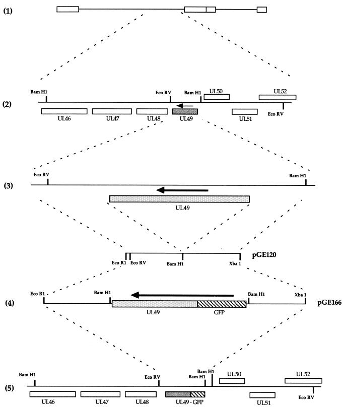
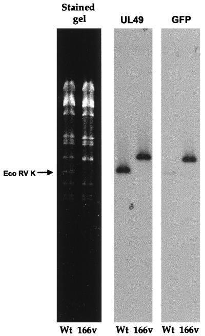
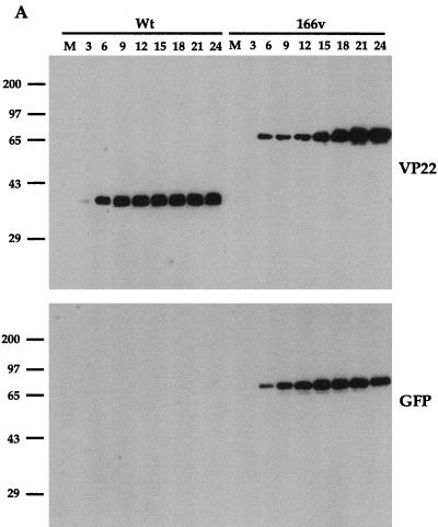
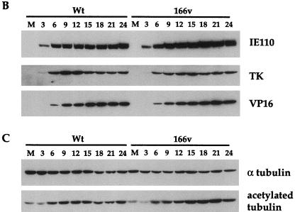
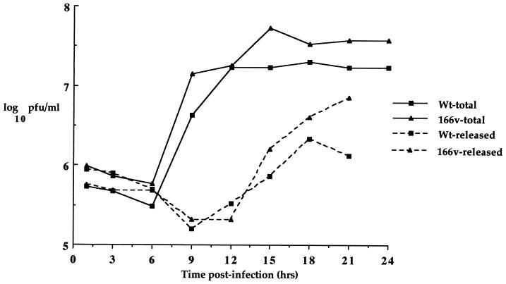
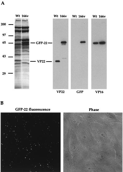
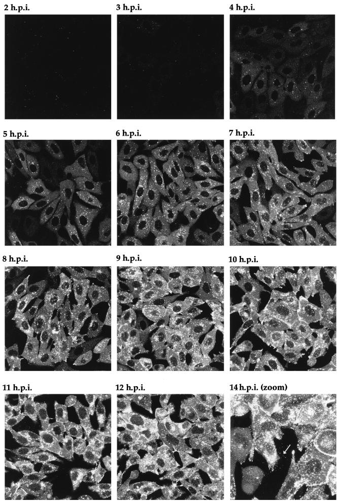
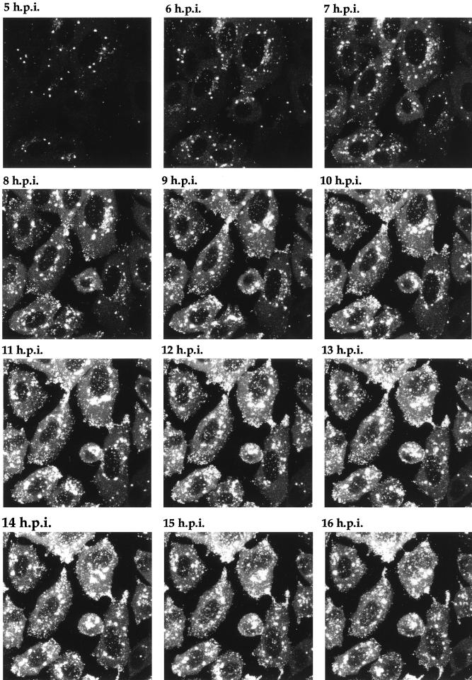
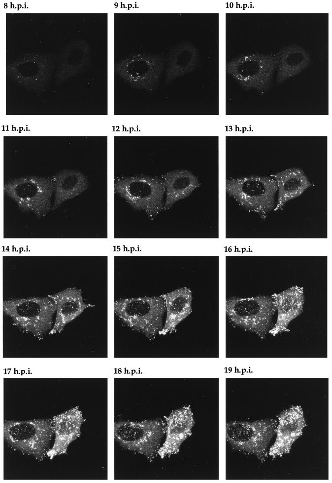
Many stages of the herpes simplex virus maturation pathway have not yet been defined. In particular, little is known about the assembly of the virion tegument compartment and its subsequent incorporation into maturing virus particles. Here we describe the construction of a herpes simplex virus type 1 (HSV-1) recombinant in which we have replaced the gene encoding a major tegument protein, VP22, with a gene expressing a green fluorescent protein (GFP)-VP22 fusion protein (GFP-22). We show that this virus has growth properties identical to those of the parental virus and that newly synthesized GFP-22 is detectable in live cells as early as 3 h postinfection. Moreover, we show that GFP-22 is incorporated into the HSV-1 virion as efficiently as VP22, resulting in particles which are visible by fluorescence microscopy. Consequently, we have used time lapse confocal microscopy to monitor GFP-22 in live-cell infection, and we present time lapse animations of GFP-22 localization throughout the virus life cycle. These animations demonstrate that GFP-22 is present in a diffuse cytoplasmic location when it is initially expressed but evolves into particulate material which travels through an exclusively cytoplasmic pathway to the cell periphery. In this way, we have for the first time visualized the trafficking of a herpesvirus structural component within live, infected cells.


Figures

FIG. 1
FIG. 1
Construction of the GFP-22-expressing virus. (1) Schematic representation of the HSV-1 genome. (2) The region of the genome which contains the UL49 gene (shaded). The arrow indicates the direction of transcription. (3) The EcoRV/BamHI fragment which contains the UL49 gene and its flanking sequences. The 400-bp sequences on either side of the UL49 gene were amplified by PCR with EcoRI and XbaI sites at either end, and a BamHI site in place of the UL49 gene (4). The EcoRI/XbaI fragment was inserted into EcoRI/XbaI-digested pSP72 to make pGE120. A GFP-VP22 BamHI cassette was inserted into the BamHI site of pGE120 to make pGE166. (5) The structure of the genome resulting from recombination of plasmid pGE166 with HSV-1 DNA.
FIG. 2
FIG. 2
Southern blotting of virus DNA confirms the presence of the GFP-22 cassette in the HSV-1 genome. Viral DNA purified from cells infected with either S17 or 166v was digested overnight with EcoRV and electrophoresed in a 0.8% agarose gel containing ethidium bromide. The gel was photographed (stained gel), transferred to a nylon membrane, and hybridized with a probe specific for the UL49 gene. The same membrane was stripped and rehybridized with a probe specific for GFP. Arrow points to the WT EcoRV K fragment.
FIG. 3
FIG. 3
The GFP-22 virus replicates as efficiently as the WT virus. Vero cells infected with either the WT virus or 166v at 10 PFU per cell were harvested every 3 h after infection, up to 24 h, as indicated above the gels. Equal amounts of total cell lysates were analyzed by SDS-PAGE followed by Western blotting with antibodies against VP22 and GFP (A); IE110, TK, and VP16 (B); or α tubulin and acetylated tubulin (C).
FIG. 3
FIG. 3
The GFP-22 virus replicates as efficiently as the WT virus. Vero cells infected with either the WT virus or 166v at 10 PFU per cell were harvested every 3 h after infection, up to 24 h, as indicated above the gels. Equal amounts of total cell lysates were analyzed by SDS-PAGE followed by Western blotting with antibodies against VP22 and GFP (A); IE110, TK, and VP16 (B); or α tubulin and acetylated tubulin (C).
FIG. 4
FIG. 4
One-step growth curves for WT and 166v viruses. Vero cells infected with either the WT virus or 166v at 10 PFU per cell were harvested every 3 h after infection up to 24 h and were titrated onto Vero cells. Total, virus yield from intra- and extracellular virus. Released, virus yield from extracellular virus.
FIG. 5
FIG. 5
Incorporation of GFP-22 into virus particles. (A) Purified WT and 166v virions were solubilized and analyzed by SDS-PAGE on a 9% acrylamide gel, followed by either Coomassie blue staining (left) or Western blotting with antibodies against VP22, GFP, or VP16 (right). VP22 and GFP-22 species are indicated. (B) 166v virions are fluorescent. Approximately 10 PFU of purified 166v virions per cell was laid onto a monolayer of Vero cells maintained at 4°C. Thirty minutes later the cells were examined live by both phase-contrast (Phase) and fluorescence (GFP-22 fluorescence) microscopy.
FIG. 6
FIG. 6
Live-cell analysis of GFP-22 localization during a high-multiplicity infection of 166v. Vero cells were infected with 166v at 10 PFU per cell and were examined every hour up to 14 h postinfection (14 h.p.i.) for GFP-22 fluorescence. The same settings for the confocal microscope were used at each time point. Extracellular fluorescent particles can be seen in the image taken at 14 h postinfection (arrow).
FIG. 7
FIG. 7
Time lapse analysis of GFP-22 trafficking in a high-multiplicity 166v infection. Vero cells were infected with 166v at 10 PFU per cell and were transferred to the heated chamber 5 h postinfection (5 h.p.i.). A single field was chosen for analysis, and images were collected every 5 min for a further 15 h. A time point representing each hour is shown, and the corresponding animation can be found at http://mc11.mcri.ac.uk/mov/figure7.mov.
FIG. 8
FIG. 8
Time lapse analysis of GFP-22 trafficking in a low-multiplicity 166v infection. Vero cells infected with 166v at 0.02 PFU per cell were transferred to the heated chamber 8 h postinfection (8 h.p.i.). A single field of cells was chosen for further analysis, and images were collected every 5 min for a further 14 h. A time point representing each hour is shown, and the corresponding animation can be found at http://mc11.mcri.ac.uk/mov/figure8.mov.

References

    1. Browne H, Bell S, Minson T, Wilson D W. An endoplasmic reticulum-retained herpes simplex virus glycoprotein H is absent from secreted virions: evidence for reenvelopment during egress. J Virol. 1996;70:4311–4316. - PMC - PubMed
    1. Campadelli-Fiume G, Farabegoli F, Di Gaeta S, Roizman B. Origin of unenveloped capsids in the cytoplasm of cells infected with herpes simplex virus 1. J Virol. 1991;65:1589–1595. - PMC - PubMed
    1. Cheung P, Banfield B W, Tufaro F. Brefeldin A arrests the maturation and egress of herpes simplex virus particles during infection. J Virol. 1991;65:1893–1904. - PMC - PubMed
    1. Dargin D. The structure and assembly of herpes viruses. In: Harris J R, Horne R W, editors. Electron microscopy of proteins. 5. Virus structure. London, United Kingdom: Academic Press; 1986. pp. 359–437.
    1. Desai P, Person S. Incorporation of the green fluorescent protein into the herpes simplex virus type 1 capsid. J Virol. 1998;72:7563–7568. - PMC - PubMed

Publication types

MeSH terms

Substances

LinkOut - more resources