. 1999 May;73(5):4181-7.
doi: 10.1128/JVI.73.5.4181-4187.1999.

Cellular tropism and viral interleukin-6 expression distinguish human herpesvirus 8 involvement in Kaposi's sarcoma, primary effusion lymphoma, and multicentric Castleman's disease

Affiliations

Cellular tropism and viral interleukin-6 expression distinguish human herpesvirus 8 involvement in Kaposi's sarcoma, primary effusion lymphoma, and multicentric Castleman's disease

K A Staskus et al. J Virol. 1999 May.

Abstract

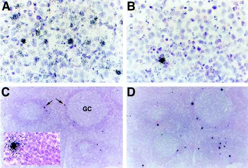
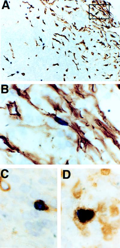
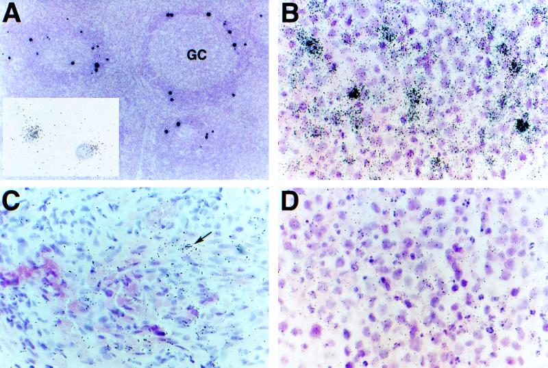
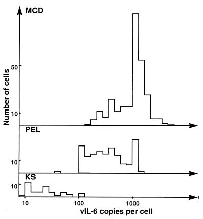
Human herpesvirus 8 (HHV-8) infection has been implicated in the etiology of Kaposi's sarcoma (KS), primary effusion lymphoma (PEL), and multicentric Castleman's disease (MCD), three diseases that frequently develop in immunocompromised, human immunodeficiency virus-positive individuals. One hypothesis that would account for different pathological manifestations of infection by the same virus is that viral genes are differentially expressed in heterogeneous cell types. To test this hypothesis, we analyzed the localization and levels of expression of two viral genes expressed in latent and lytic infections and the viral homologue of interleukin-6 (vIL-6). We show that PEL parallels KS in the pattern of latent and lytic cycle viral gene expression but that the predominant infected cell type is a B cell. We also show that MCD differs from KS not only in the infected cell type (B-cell and T-cell lineage) but also in the pattern of viral gene expression. Only a few cells in the lesion are infected and all of these cells express lytic-cycle genes. Of possibly greater significance is the fact that in a comparison of KS, PEL, and MCD, we found dramatic differences in the levels of expression of vIL-6. Interleukin-6 is a B-cell growth and differentiation factor whose altered expression has been linked to plasma cell abnormalities, as well as myeloid and lymphoid malignancies. Our findings support the hypothesis that HHV-8 plays an important role in the pathogenesis of PEL and MCD, in which vIL-6 acts as an autocrine or paracrine factor in the lymphoproliferative processes common to both.


Figures

FIG. 1
FIG. 1
In situ localization of HHV-8-infected cells in PEL and MCD. Thin sections of PEL (A and B) and MCD (C and D) specimens were hybridized with 35S-labeled riboprobes specific for HHV-8 genes expressed during latency (T0.7) and productive infection (nut-1). PEL displays a hybridization pattern similar to that which we have previously reported for KS: T0.7 (A, 3-day exposure) is expressed in a majority of cells of the effusion and to various levels across the population of infected cells (visualized as various amounts of silver grains that have developed in the photographic emulsion coating the specimen), whereas nut-1, which potentially indicates lytic infection, is transcribed in only a few of the total infected cells (B, 18-h exposure). Hybridizations to MCD reveal that, relative to PEL, few cells express T0.7; that expression is to relatively high levels (collections of silver grains over individual cells seen as black dots on this overexposed slide at this low magnification; arrows); and that these cells are localized mainly to the “onionskin” perifollicular collections of lymphocytes surrounding the germinal centers (GC) (C, 3-day exposure). Unlike KS and PEL, the pattern of nut-1 hybridization, with respect to location and numbers of positive cells, is similar to that of T0.7 in the same tissue (D, 18-h exposure). Like KS and PEL, these cells express nut-1 to much higher levels than T0.7. Higher magnifications of panels C (inset) and D do not reveal a significant number of additional positive cells that cannot be seen at this magnification. Counterstaining was done with hematoxylin and eosin.
FIG. 2
FIG. 2
Identification of HHV-8-infected cells in MCD. Combined ISH with a digoxigenin-labeled nut-1 riboprobe (dark purple nuclei) and IHC with a monoclonal antibody to human CD34 (brown peroxidase reaction product) reveals the presence of an infected spindle-shaped cell, similar to the KS tumor cell, in a collection of CD34-staining cells in the subcapsular sinus of a lymph node with MCD seen at low (A, boxed area, arrow, upper right) and high (B) magnifications. Other infected cells (A, arrows, center and lower left) in the perifollicular lymphocyte layer do not express CD34. Colocalization of the digoxigenin-labeled nut-1 riboprobe and antibody to the human lambda light chain (C) or CD3 (D) shows that some of these infected CD34-negative cells are plasma cells and T cells, respectively. Counterstaining was done briefly with hematoxylin.
FIG. 3
FIG. 3
vIL-6 expression in KS, PEL, and MCD. Hybridization of MCD with a 35S-labeled vIL-6 riboprobe reveals high levels of transcripts in cells localized mainly to the perifollicular lymphocyte layer of the specimen (A, 3-day exposure). These cells are similar in quantity and location to those transcribing T0.7 and nut-1 RNAs in subjacent sections (Fig. 1C and D) and double ISH with 35S-labeled vIL-6 and digoxigenin-labeled nut-1 riboprobes show colocalization to the same cells (A, inset, 2-h exposure). A higher magnification of this overexposed slide does not reveal additional vIL-6-positive cells. Hybridization of 35S-labeled vIL-6 to PEL shows that, like hybridization for T0.7 in PEL, a majority of cells are transcribing this gene and to various levels within the population (B, 3-day exposure) but to a visibly lesser extent than in MCD. KS expresses the least vIL-6 (C, 7-day exposure) with an infrequent cell containing few transcripts (arrow) among a majority of T0.7-containing cells (hybridization not shown). Control hybridization of a 35S-labeled riboprobe for human IL-6 to PEL shows lack of cross-reactivity (D, 3-day exposure). Counterstaining was done with hematoxylin and eosin.
FIG. 4
FIG. 4
Frequency distribution of vIL-6 copy number in MCD, PEL, and KS. The number of copies of vIL-6 RNA in individual infected cells was determined by using computerized image analysis to count grains over randomly selected cells. Grain counts were converted to copy numbers as previously described (6). MCD: range, 270 to 7,942 copies per cell; median, 1,415 copies per cell; mean, 1,790 copies per cell; 281 cells counted. PEL: range, 66 to 2,371 copies per cell; median, 555 copies per cell; mean, 648 copies per cell; 168 cells counted. KS: range, 9 to 156 copies per cell; median, 46 copies per cell; mean, 40 copies per cell; 44 cells counted.

Similar articles

Cited by

References

    1. Ansari M Q, Dawson D B, Nador R, Rutherford C, Schneider N R, Latimer M J, Ricker L, Knowles D M, McKenna R W. Primary body cavity-based AIDS-related lymphomas. Am J Clin Pathol. 1996;105:141–143. - PubMed
    1. Arcone R, Pucci P, Zappacosta F, Fontaine V, Malorni A, Marino G, Ciliberto G. Single-step purification and structural characterization of human interleukin-6 produced in Escherichia coli from a T7 RNA polymerase expression vector. Eur J Biochem. 1991;198:541–547. - PubMed
    1. Barozzi P, Luppi M, Masini L, Marasca R, Savarino M, Morselli M, Ferrari M G, Bevini M, Bonacorsi G, Torelli G. Lymphotropic herpes virus (EBV, HHV-6, HHV-8) DNA sequences in HIV negative Castleman’s disease. J Clin Pathol. 1996;49:M232–M235. - PMC - PubMed
    1. Burger R, Wendler J, Antoni K, Helm G, Kalden J R, Gramatzki M. Interleukin-6 production in B-cell neoplasias and Castleman’s disease: evidence for an additional paracrine loop. Ann Hematol. 1994;69:25–31. - PubMed
    1. Castleman B, Iverson L, Menendez V P. Localized mediastinal lymph-node hyperplasia resembling thymoma. Cancer. 1956;9:822–830. - PubMed

Publication types