. 1999 May;73(5):4220-9.
doi: 10.1128/JVI.73.5.4220-4229.1999.

CCAAT displacement protein binds to and negatively regulates human papillomavirus type 6 E6, E7, and E1 promoters

Affiliations

CCAAT displacement protein binds to and negatively regulates human papillomavirus type 6 E6, E7, and E1 promoters

W Ai et al. J Virol. 1999 May.

Abstract

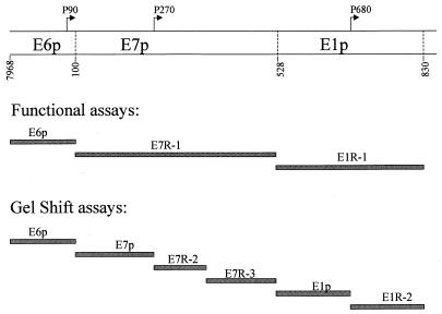
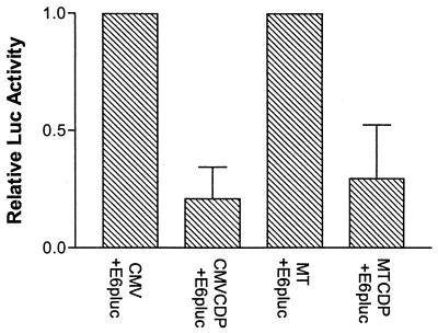
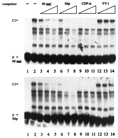
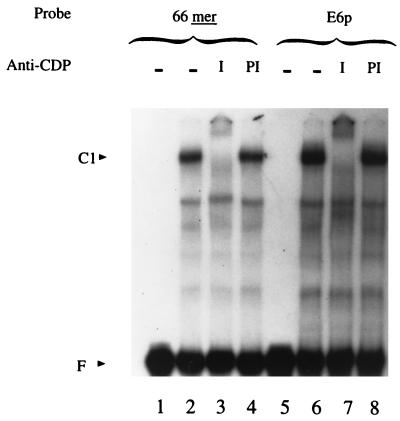
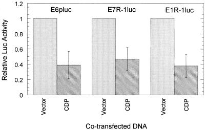
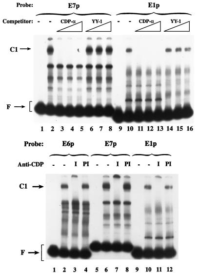
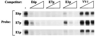
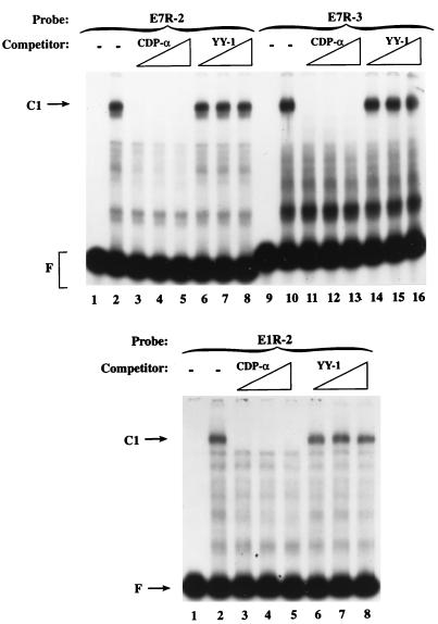
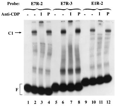
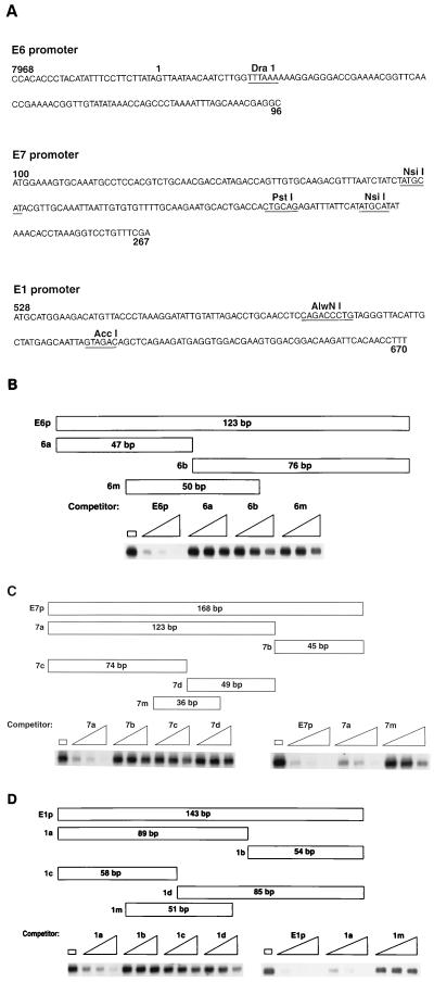
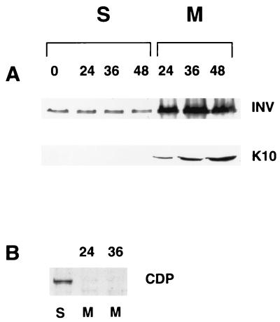
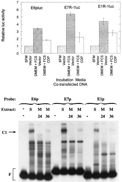
Expression of human papillomavirus genes increases as the target cell, the keratinocyte, differentiates. CCAAT displacement protein (CDP) is a cellular protein which has been shown in other cell types to negatively regulate gene expression in undifferentiated cells but not in differentiated cells. We have previously shown that a 66-bp purine-thymidine-rich sequence (the 66-mer) binds CDP and negatively regulates the human papillomavirus type 6 (HPV-6) E6 promoter (S. Pattison, D. G. Skalnik, and A. Roman, J. Virol. 71:2013-2022, 1997). Cotransfection experiments with a plasmid expressing luciferase from the HPV-6 E6, E7, or E1 regulatory region and a plasmid carrying the CDP gene indicate that CDP represses transcription from all three HPV-6 promoters. Using electrophoretic mobility shift assays (EMSAs), we have shown that CDP binds HPV-6 both upstream and downstream of the E6, E7, and E1 transcription initiation start sites. Furthermore, when keratinocytes were induced to differentiate, all three promoter activities increased. Consistent with this, immunoblotting and EMSAs revealed that endogenous nucleus CDP and, correspondingly, DNA binding activity decreased when keratinocytes were induced to differentiate. The elevated promoter activities were abrogated by exogenously transfected CDP. Our data demonstrate that CDP fulfills the requirement of a differentiation-dependent negative regulator that could tie the HPV life cycle to keratinocyte differentiation.


Figures

FIG. 1
FIG. 1
Location of fragments used in functional assays and EMSAs. The top panel shows the three HPV-6W50 early promoter regulatory regions. The arrows indicate E6, E7, and E1 transcriptional initiation start sites. The dashed lines indicate E6, E7, and E1 translational start sites, respectively. The middle panel shows the three regulatory regions used in the functional studies. E6p extends from nt 7968 within the LCR to nt 96, 4 bp upstream of the E6 translational start site. E7R-1 extends from the E6 translational start site to the E7 translational start site; E1R-1 extends from the E7 translational start site to the E1 translational start site. In the bottom panel, the fragments used in EMSAs are shown. E7R-1 was divided into E7p, E7R-2, and E7R-3; E1R-1 was divided into E1p and E1R-2. Sequences used to amplify fragments are listed in Table 1. The cloning strategy is provided in Materials and Methods.
FIG. 2
FIG. 2
CDP negatively regulates the HPV-6 E6 promoter. E6pluc was cotransfected with either empty vector carrying the promoter (CMV or MT) or the CDP expression plasmid (CMVCDP or MTCDP) into human primary keratinocytes, along with the internal control CMVβ-gal. Forty to 48 h later, the cells were harvested and the luciferase (Luc) activities were assayed as described in Materials and Methods. The standardized luciferase activity obtained for the E6pluc parental plasmid in the presence of empty vector (CMV or MT) was set to 1.0. The average ± standard deviation for three experiments is shown.
FIG. 3
FIG. 3
CDP binds to the HPV-6 E6 promoter in addition to the 66-mer. EMSAs were carried out with the 66-mer (upper panel) and the E6 promoter (lower panel) as probes. Lane 1 shows each free probe; lane 2 shows the result of the binding assay in the absence of competitors. The remaining lanes show competition assays in the presence of a 50-, 100-, or 500-fold molar excess of the indicated competitors.
FIG. 4
FIG. 4
Confirmation of CDP binding to the E6 promoter by using anti-CDP antiserum. The 66-mer and the E6 promoter (E6p) probes were used in EMSAs. Lanes: 1 and 5, free probes (F); 2 and 6, assays performed in the absence of serum; 3 and 7, assays performed in the presence of anti-CDP antiserum (I); 4 and 8, assays performed in the presence of preimmune serum (PI).
FIG. 5
FIG. 5
CDP negatively regulates E6, E7, and E1 promoter activities. Cotransfection assays were carried out with E6luc, E7R-1luc, and E1R-1luc with either a plasmid carrying the CMV promoter (vector) or CMV-driven CDP (CDP) in human primary keratinocytes. The luciferase activity was normalized as for Fig. 2. The average ± standard deviation for a representative experiment, conducted in duplicate, is shown.
FIG. 6
FIG. 6
CDP binds to the E7 and E1 promoters. (Top) EMSAs were performed with the E7 promoter (E7p) or the E1 promoter (E1p) as a probe. Lanes 1 and 9 show each free probe. Lanes 2 and 10 show assays in the absence of competitors (−). The remaining lanes show competition assays in the presence of a 20-, 50-, or 200-fold molar excess of the indicated oligonucleotide. (Bottom) The E6 promoter (E6p), the E7 promoter (E7p), and the E1 promoter (E1p) probes were used in EMSAs. Lanes 1, 5, and 9 show the free probes (F). The remaining lanes show assays performed in the absence of serum (−), in the presence of anti-CDP antiserum (I), or in the presence of preimmune serum (PI).
FIG. 7
FIG. 7
CDP binding affinities for E6, E7, and E1 promoters. The E6 promoter (E6p), E7 promoter (E7p), and E1 promoter (E1p) were used as probes in EMSAs, in the absence (−) or presence of a 20-, 50-, or 200-fold molar excess of the indicated competitors.
FIG. 8
FIG. 8
CDP binds to the E7 and E1 downstream fragments. EMSAs were carried out with E7R-2, E7R-3, or E1R-2 as a probe. Lanes 1 and 9 in the top panel and lane 1 in the bottom panel show each free probe (F). Lanes 2 and 10 in the top panel and lane 2 in the bottom panel show assays in the absence of competitors. The remaining lanes show competition assays in the presence of a 20-, 50-, or 200-fold molar excess of the indicated oligonucleotide.
FIG. 9
FIG. 9
Confirmation of CDP binding to the E7 and E1 downstream fragments by using anti-CDP antiserum. E7R-2, E7R-3, or E1R-2 was used as a probe in EMSAs in the absence of antiserum (−), in the presence of anti-CDP antiserum (I), or in the presence of preimmune serum (PI). F, free probes.
FIG. 10
FIG. 10
At least two regions within the E6, E7 and E1 promoters are required for CDP to bind. (A) Sequences of the E6, E7, and E1 promoters. Numbers indicate the nucleotide positions in the HPV-6aW50 genome. Underlined sequences indicate the restriction sites, and the corresponding restriction enzymes are listed above the recognition sequences. (B) E6 promoter subfragment competitors and EMSAs. (Top) The diagram depicts the relationship of the subfragments used in the EMSAs. The length of each fragment is marked within each fragment (see Materials and Methods for detailed information about each fragment). (Bottom) EMSAs were carried out with the E6 promoter as a probe in the presence of no competitor or a 20-, 50-, or 200-fold molar excess of the indicated competitors. (C) E7 promoter subfragment competitors and EMSAs. (Top) Diagram of the fragments used in the EMSAs. (Bottom) EMSAs were conducted as for panel B with the E7 promoter as a probe and E7p, 7a, 7b, 7c, 7d, and 7m as unlabeled competitors. (D) E1 promoter subfragment competitors and EMSAs. (Top) Diagram of the subfragments used in the EMSAs. (Bottom) EMSAs were conducted as for panel B with the E1 promoter as a probe and E1p, 1a, 1b, 1c, 1d, and 1m as unlabeled competitors.
FIG. 11
FIG. 11
Keratinocyte markers indicate cells suspended in methylcellulose are differentiated. (A) Fifty micrograms of whole-cell extract from cells suspended in methylcellulose was separated on polyacrylamide gels, and the proteins were transferred to nitrocellulose and probed with antibodies to involucrin (INV) or keratin 10 (K10). (B) Fifty micrograms of the nuclear extract used in EMSAs was similarly analyzed for the presence of CDP. S, Cells grown in SFM for the indicated time in hours (36 h in panel B); M, cells grown in 1.5% methylcellulose for the indicated time in hours.
FIG. 12
FIG. 12
Increased HPV-6 early promoter activities correlate with the decreased CDP binding activity in differentiated keratinocytes. (Top) Cotransfection experiments were performed as in Fig. 5. The luciferase activities obtained in the differentiation media with the empty vector or CDP expression vector were compared to luciferase activities obtained with the regulatory region cotransfected with the empty vector (CMV) and maintained in SFM. FCS, fetal calf serum. The results represent the average ± standard deviation of two experiments each conducted in duplicate. (Bottom) EMSAs were performed with the E6 promoter (E6p), E7 promoter (E7p), or E1 promoter (E1p) as a probe and nuclear extracts from keratinocytes growing in SFM (S) or from keratinocytes suspended in methylcellulose for 24 h (M24) or 36 h (M36). F, free probes.

References

    1. Andersen B, Hariri A, Pittelkow M R, Rosenfeld M G. Characterization of Skn-1a/i POU domain factors and linkage to papillomavirus gene expression. J Biol Chem. 1997;272:15905–15913. - PubMed
    1. Andres V, Chiara M D, Mahdavi V. A new bipartite DNA-binding domain: cooperative interaction between the cut repeat and homeo domain of the cut homeo proteins. Genes Dev. 1994;8:245–257. - PubMed
    1. Andres V, Nadal-Ginard B, Mahdavi V. Clox, a mammalian homeobox gene related to Drosophila cut, encodes DNA-binding regulatory proteins differentially expressed during development. Development. 1992;116:321–334. - PubMed
    1. Apt D, Watts R M, Suske G, Bernard H U. High Sp1/Sp3 ratios in epithelial cells during epithelial differentiation and cellular transformation correlate with the activation of the HPV-16 promoter. Virology. 1996;224:281–291. - PubMed
    1. Aufiero B, Neufeld E J, Orkin S H. Sequence-specific DNA binding of individual cut repeats of the human CCAAT displacement/cut homeodomain protein. Proc Natl Acad Sci USA. 1994;91:7757–7761. - PMC - PubMed

Publication types

Associated data

LinkOut - more resources