. 1999 Apr;10(4):833-45.
doi: 10.1091/mbc.10.4.833.

Cdc25 inhibited in vivo and in vitro by checkpoint kinases Cds1 and Chk1

Affiliations
Free PMC article

Cdc25 inhibited in vivo and in vitro by checkpoint kinases Cds1 and Chk1

B Furnari et al. Mol Biol Cell. 1999 Apr.
Free PMC article

Abstract

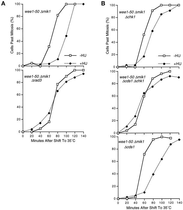
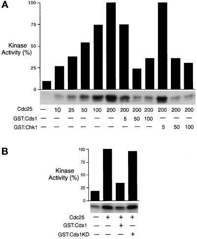
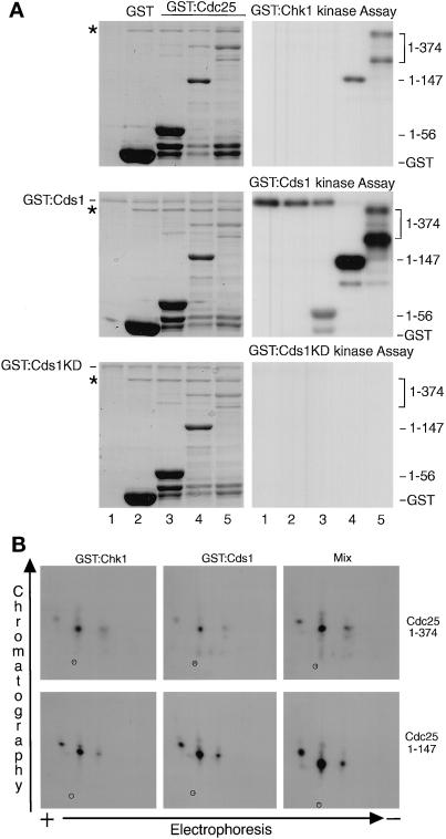
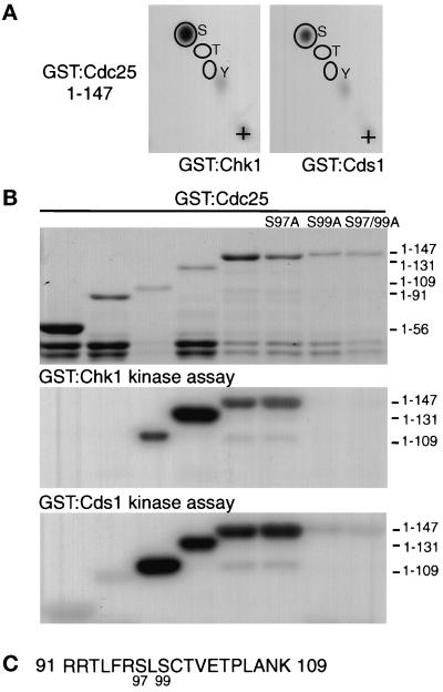
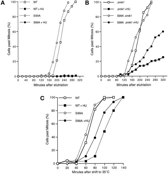
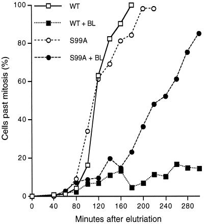
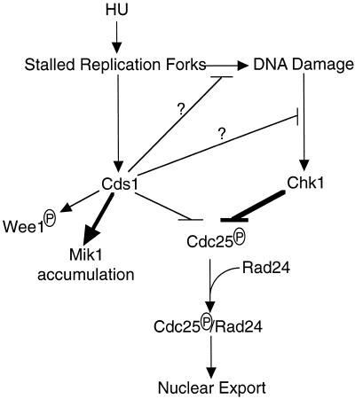
In the fission yeast Schizosaccharomyces pombe, the protein kinase Cds1 is activated by the S-M replication checkpoint that prevents mitosis when DNA is incompletely replicated. Cds1 is proposed to regulate Wee1 and Mik1, two tyrosine kinases that inhibit the mitotic kinase Cdc2. Here, we present evidence from in vivo and in vitro studies, which indicates that Cds1 also inhibits Cdc25, the phosphatase that activates Cdc2. In an in vivo assay that measures the rate at which Cdc25 catalyzes mitosis, Cds1 contributed to a mitotic delay imposed by the S-M replication checkpoint. Cds1 also inhibited Cdc25-dependent activation of Cdc2 in vitro. Chk1, a protein kinase that is required for the G2-M damage checkpoint that prevents mitosis while DNA is being repaired, also inhibited Cdc25 in the in vitro assay. In vitro, Cds1 and Chk1 phosphorylated Cdc25 predominantly on serine-99. The Cdc25 alanine-99 mutation partially impaired the S-M replication and G2-M damage checkpoints in vivo. Thus, Cds1 and Chk1 seem to act in different checkpoint responses to regulate Cdc25 by similar mechanisms.


Figures

Figure 1
Figure 1
Rad3, Cds1, and Chk1 contribute to the HU-induced delay of mitosis after inactivation of Wee1 and Mik1. (A) Cells having rad3+ or Δrad3 alleles in the wee1-50 Δmik1 background were grown to midlog phase at the permissive temperature of 25°C and synchronized in early G2 phase by centrifugal elutriation. HU was added at a concentration of 12 mM to one-half of each culture at 40 min. The culture temperature was shifted to the restrictive temperature of 35°C after the cells had divided once. HU caused a delay of ∼40 min in the rad3+ strain (top), whereas no delay was observed in the Δrad3 strain (bottom). The activity of Cdc25 determines the rate of mitotic onset after inactivation of Wee1 and Mik1. Rad3 is required for the replication checkpoint; thus these data indicate that the checkpoint inhibits the function of Cdc25 in vivo. (B) An experiment was performed to determine the contributions of Cds1 and Chk1 in the HU-induced checkpoint delay that is observed after inactivation of Wee1 and Mik1. The checkpoint delay was abolished in a Δcds1 Δchk1 background (middle). The duration of the delay was ∼20 min in Δchk1 cells (top), compared with an ∼40-min delay in a cds1+ chk1+ background. Thus, Cds1 contributes to the HU-induced inhibition of Cdc25 function in vivo. The delay appeared to be fully restored in the Δcds1 cells (bottom), which indicates that Chk1 makes a major contribution to the HU-induced mitotic delay. However, it is possible that the Δcds1 mutation leads to greater activation of Chk1, thereby appearing to exaggerate the contribution of Chk1 (Lindsay et al., 1998).
Figure 2
Figure 2
Cds1 and Chk1 inactivate Cdc25 in vitro. (A) Hexahistidine-tagged S. pombe Cdc25 was expressed and purified from insect cells and used to activate Cdc2/cyclin-B1 purified from HeLa cells arrested in early S phase with thymidine. Antibodies to human cyclin-B1 were used to purify Cdc2/cyclin-B1 that was assayed by its ability to transfer [γ-32P]ATP to histone H1. Cdc25 was purified from 1 × 10⁷ cells and suspended in 1 ml. Numbers in the Cdc25 row refer to the volume of Cdc25 preparation added to the reaction (see MATERIALS AND METHODS). Cdc25 caused a dose-dependent activation of Cdc2/cyclin-B. GST–Cds1 and GST–Chk1 were purified from fission yeast and used to phosphorylate Cdc25 before incubation with Cdc2/cyclin-B (see MATERIALS AND METHODS). Numbers in the GST–Cds1 and GST–Chk1 rows refer to the volume added of a 660-μl preparation made from 20 OD600 of cells. GST–Cds1 and GST–Chk1 caused a dose-dependent inhibition of Cdc25. The graph presents phosphorimager analysis of the autoradiogram shown below the graph. (B) The Cdc25 assay described above was repeated with GST–Cds1KD (GST–Cds1D312E), a kinase-inactive form of Cds1. Relative to active GST–Cds1, GST–Cds1KD only modestly decreased activation of Cdc2/cyclin-B by Cdc25. Coomassie blue staining confirmed that equal amounts of GST–Cds1 and GST–Cds1KD were added to these reactions (see Figure 3).
Figure 3
Figure 3
Cds1 and Chk1 phosphorylate the NH2-terminal domain of Cdc25 to generate very similar phosphopeptide maps. (A) GST alone (lane 2) or GST fusion proteins containing amino acids 1–56, 1–147, or 1–374 of fission yeast Cdc25 (lanes 3–5, respectively) were expressed and purified from bacteria. The proteins were phosphorylated with GST–Chk1 or GST–Cds1 purified from fission yeast. Left, Coomassie blue stain analysis of protein gels is shown. Fusion proteins, degradation products, and GST–Cds1 are visible. There was insufficient GST–Chk1 to detect by Coomassie blue staining, but it was readily detected by immunoblotting (our unpublished observations). A major contaminating protein (*) from bacteria is indicated. Right, autoradiograms are shown. The 1–147 and 1–374 fusion proteins were effectively phosphorylated by the protein kinases, whereas unfused GST or the 1–56 fusion proteins were phosphorylated weakly or not at all. A 1–374 degradation product containing ∼200 amino acids from Cdc25 was also phosphorylated. Phosphorylated GST–Cds1 was also detected, predominantly in lanes 1–3. These reactions did not contain a preferred substrate for GST–Cds1, perhaps accounting for the increased autophosphorylation in these samples. GST–Cds1KD (GST–Cds1D312E), the kinase-inactive form of Cds1, was unable to phosphorylate itself or the GST–Cdc25 fusion proteins. Coomassie blue staining confirmed that equal amounts of GST–Cds1 and GST–Cds1KD were used in these studies. The same protein preparations were used for the experiment shown in Figure 2B. (B) Two-dimensional tryptic phosphopeptide maps of GST–Cdc251–374 and GST–Cdc251–147 phosphorylated by GST–Cds1 (middle) or GST–Chk1 (left) appear very similar. Experiments in which the Chk1 and Cds1 reactions of GST–Cdc251–374 or GST–Cdc251–147 (right) were mixed confirm the similarity of the maps.
Figure 4
Figure 4
Cds1 and Chk1 phosphorylate serine-99 of Cdc25. (A) Two-dimensional phosphoamino acid analysis revealed that GST–Cds1 (right) and GST–Chk1 (left) phosphorylated GST–Cdc251–147 exclusively on serine. (B) Various regions of Cdc25 were expressed as GST fusion proteins in bacteria and tested as substrates for GST–Cds1 (bottom) or GST–Chk1 (middle). A Coomassie blue stain of the protein gel indicates the relative amounts and positions of fusion proteins and degradation products (top). Autoradiograms showed that the 1–109 construct was phosphorylated by both kinases, whereas the 1–91 construct was not phosphorylated. This result indicated that the major phosphorylation site is located in the 91–109 region of Cdc25. Mutation of serine-97 to alanine (S97A) did not change phosphorylation of the 1–147 construct, whereas the S99A and S97/99A forms of the 1–147 construct were not phosphorylated. Thus, serine-99 appears to be the major site of phosphorylation. (C) Sequence of the 91–109 region of Cdc25 is shown.
Figure 5
Figure 5
The cdc25-S99A mutation impairs the S–M checkpoint. (A and B) Plasmids containing cdc25+ (WT) or cdc25-S99A (S99A) were integrated in cdc25-22 or cdc25-22 mik1::LEU2 cells. Synchronous cultures of cells in early S phase were produced by pretreatment with HU (12 mM) for 120 min at 25°C followed by centrifugal elutriation in the presence of HU. The cells were then washed and resuspended in medium with or without HU. Mock-treated cells underwent mitosis with similar kinetics. (A) The WT and S99A cells were unable to divide during the course of the experiment, indicative of a checkpoint arrest. (B) Approximately 25% of the Δmik1 cells underwent mitosis during the course of the experiment. This number was increased to ∼60% in S99A Δmik1 cells. These findings indicate that the Δmik1 mutation impairs the checkpoint and that this effect is exacerbated by the S99A mutation. (C) An experiment was performed to determine the consequence of the S99A mutation in the HU-induced checkpoint delay that is observed after inactivation of Wee1 and Mik1. The cdc25+ (WT) or cdc25-S99A (S99A) plasmids were integrated into Δcdc25 wee1-50 Δmik1 cells. The experiment was performed as described in Figure 1. HU caused a delay of ∼40 min in the cdc25+ strain, whereas only an ∼20-min delay of mitosis was observed in the cdc25-S99A strain. These data suggest that the S99A mutation impaired but did not abolish the regulation of Cdc25 in response to HU.
Figure 6
Figure 6
The cdc25-S99A mutation impairs the DNA damage checkpoint. Plasmids containing cdc25+ (WT) or cdc25-S99A (S99A) were integrated in cdc25-22 cells. Synchronous cultures of cells in early G2 phase were produced by centrifugal elutriation and exposed to the radiomimetic drug bleomycin (BL) at a concentration of 5 mU/ml or mock treated. Mock-treated cells underwent mitosis with similar kinetics. Most of the WT cells (∼80%) were unable to divide during the course of the experiment, indicative of a checkpoint arrest. In contrast, all but ∼15% of the S99A mutant cells divided, indicative of a checkpoint failure.
Figure 7
Figure 7
Model of replication and repair checkpoint pathways in fission yeast. See text for discussion of model.

References

    1. Bentley NJ, Holtzman DA, Flaggs G, Keegan KS, DeMaggio A, Ford JC, Hoekstra M, Carr AM. The Schizosaccharomyces pombe rad3 checkpoint gene. EMBO J. 1996;15:6641–6651. - PMC - PubMed
    1. Blasina A, Paegle S, McGowan CH. The role of inhibitory phosphorylation of CDC2 following DNA replication block and radiation-induced damage in human cells. Mol Biol Cell. 1997;8:1013–1023. - PMC - PubMed
    1. Blasina A, Van de Weyer I, Laus MC, Luyten WHML, Parker AE, McGowan CH. A human homolog of the checkpoint kinase Cds1 directly inhibits Cdc25. Curr Biol. 1999;9:1–10. - PubMed
    1. Boddy MN, Furnari B, Mondesert O, Russell P. Replication checkpoint enforced by kinases Cds1 and Chk1. Science. 1998;280:909–912. - PubMed
    1. Booher RN, Alfa CE, Hyams JS, Beach DH. The fission yeast cdc2/cdc13/suc1 protein kinase: regulation of catalytic activity and nuclear localization. Cell. 1989;58:485–497. - PubMed

Publication types

MeSH terms