. 1999 Apr;10(4):1043-59.
doi: 10.1091/mbc.10.4.1043.

Two endoplasmic reticulum (ER) membrane proteins that facilitate ER-to-Golgi transport of glycosylphosphatidylinositol-anchored proteins

Affiliations
Free PMC article

Two endoplasmic reticulum (ER) membrane proteins that facilitate ER-to-Golgi transport of glycosylphosphatidylinositol-anchored proteins

W P Barz et al. Mol Biol Cell. 1999 Apr.
Free PMC article

Abstract

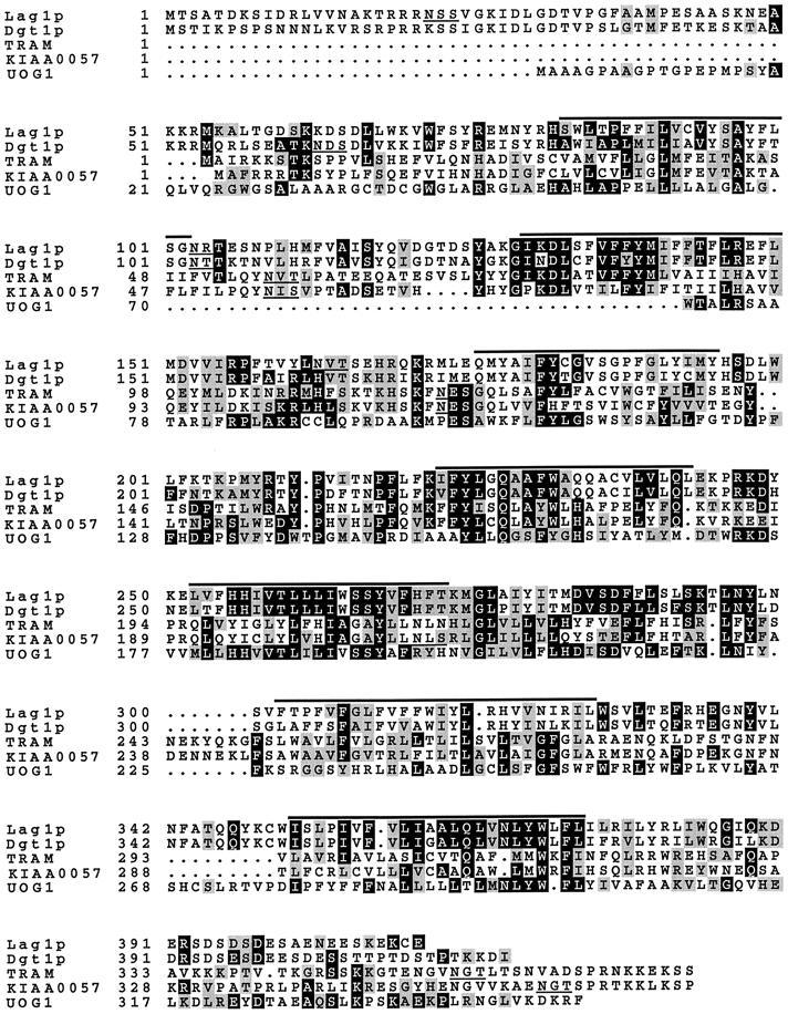
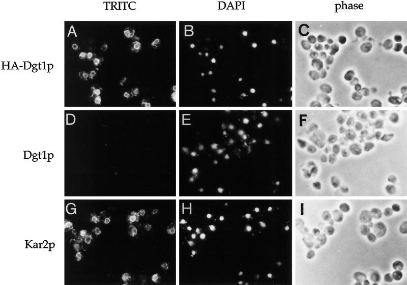
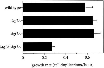
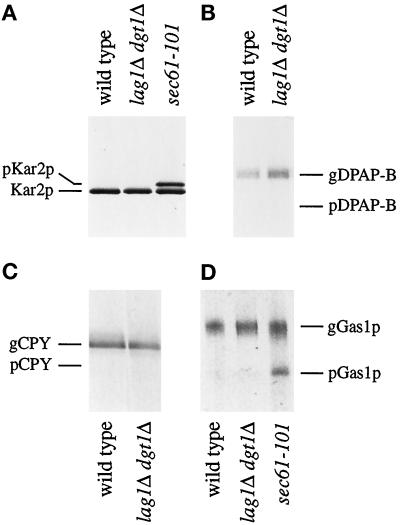
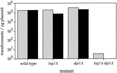
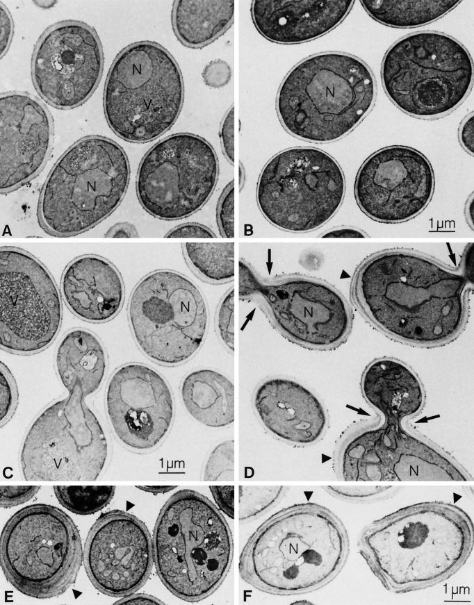
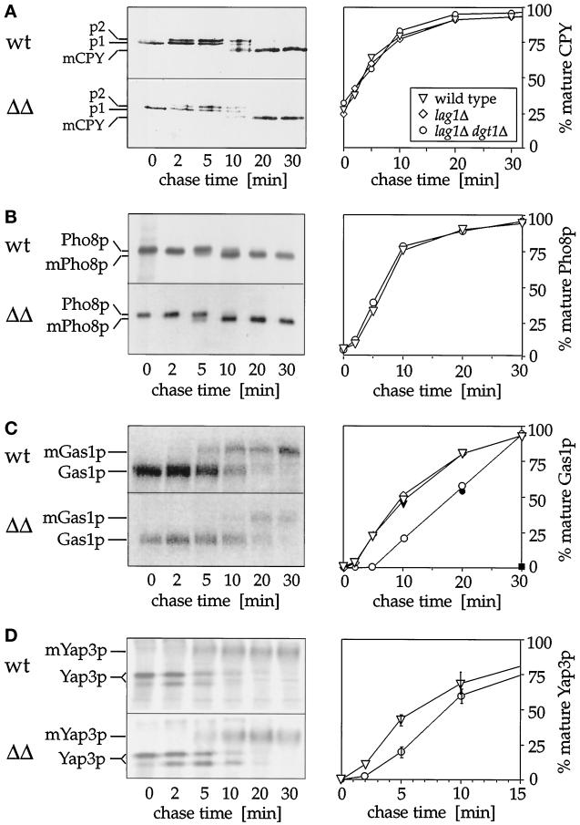
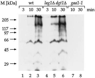
Many eukaryotic cell surface proteins are anchored in the lipid bilayer through glycosylphosphatidylinositol (GPI). GPI anchors are covalently attached in the endoplasmic reticulum (ER). The modified proteins are then transported through the secretory pathway to the cell surface. We have identified two genes in Saccharomyces cerevisiae, LAG1 and a novel gene termed DGT1 (for "delayed GPI-anchored protein transport"), encoding structurally related proteins with multiple membrane-spanning domains. Both proteins are localized to the ER, as demonstrated by immunofluorescence microscopy. Deletion of either gene caused no detectable phenotype, whereas lag1Delta dgt1Delta cells displayed growth defects and a significant delay in ER-to-Golgi transport of GPI-anchored proteins, suggesting that LAG1 and DGT1 encode functionally redundant or overlapping proteins. The rate of GPI anchor attachment was not affected, nor was the transport rate of several non-GPI-anchored proteins. Consistent with a role of Lag1p and Dgt1p in GPI-anchored protein transport, lag1Delta dgt1Delta cells deposit abnormal, multilayered cell walls. Both proteins have significant sequence similarity to TRAM, a mammalian membrane protein thought to be involved in protein translocation across the ER membrane. In vivo translocation studies, however, did not detect any defects in protein translocation in lag1Delta dgt1Delta cells, suggesting that neither yeast gene plays a role in this process. Instead, we propose that Lag1p and Dgt1p facilitate efficient ER-to-Golgi transport of GPI-anchored proteins.


Figures

Figure 1
Figure 1
Multiple alignment of the predicted amino acid sequence of TRAM homologues. The alignment includes the deduced protein sequence of S. cerevisiae LAG1 (D’mello et al. 1994), S. cerevisiae YKL008c (termed DGT1 in this work), human TRAM (Görlich et al. 1992), a human TRAM homologue (KIAA0057; Nomura et al. 1994), and human UOG-1. The alignment was generated using the PileUp (Genetics Computer Group, Madison, WI) and BOXSHADE (Institute for Animal Health, Surrey, United Kingdom) programs. Gaps that were inserted during the alignment are denoted by dots. Potential membrane-spanning domains were predicted using four different algorithms: PepPlot (Kyte and Doolittle, 1982), TMAP (Persson and Argos, 1994), TMpred (Hofmann and Stoffel, 1993), and TopPred2 (von Heijne, 1992). The positions of these putative transmembrane domains are indicated by black bars above the alignment. N-linked glycosylation consensus sequences are underlined. Black boxes indicate identical residues, whereas gray boxes show amino acid similarity.
Figure 2
Figure 2
Immunofluorescence localization of Dgt1p to the ER. lag1Δ dgt1Δ cells harboring an HA-tagged (WBY739) or an untagged (WBY743) version of DGT1 were fixed and processed for immunofluorescence using monoclonal anti-HA (A–F) or polyclonal anti-Kar2p (G–I) antibodies followed by secondary decoration with TRITC-conjugated anti-mouse or anti-rabbit IgG. Panels show the staining pattern of TRITC to visualize HA-Dgt1p and Kar2p localization. DNA was stained with DAPI to indicate nuclei, and cells were visualized by light microscopy (phase) to observe cell morphology.
Figure 3
Figure 3
Lag1p and Dgt1p are important for growth. Growth rates in YPD medium of wild type and strains in which the chromosomal copies of LAG1 DGT1 or both LAG1 and DGT1 have been disrupted are shown. Growth rates were determined at 30°C as described in MATERIALS AND METHODS. Values are the average of four to six measurements; error bars indicate SD. The growth defect was observed in different rich and minimal media containing either glucose or galactose as the carbon source. When growth rates were studied on YPD at various temperatures between 15 and 40°C, lag1Δ dgt1Δ cells grew drastically slower at all temperatures tested. The growth defect was more pronounced above 35°C, indicating a slightly increased temperature sensitivity. Although lag1Δ dgt1Δ cells formed tiny colonies at 37°C, no growth was observed at 39–40°C, at which wild-type, lag1Δ, and dgt1Δ single mutant cells grew normally.
Figure 4
Figure 4
Protein translocation across the ER membrane does not require LAG1 and DGT1. Cells of W303-1B (wild type), WBY616 (lag1Δ dgt1Δ), or DNY116 (sec61-101) were pulse labeled with [35S]methionine and [35S]cysteine for 5 min. Cell lysates were prepared, and immunoprecipitations were performed using polyclonal antibodies against the following proteins: (A) Kar2p, (B) dipeptidyl aminopeptidase B (DPAP B), (C) CPY, and (D) the GPI-anchored cell surface protein Gas1p. Eluates were subjected to SDS-PAGE followed by fluorography. Cytosolic precursor (p) and ER-glycosylated (g) forms of each protein are indicated.
Figure 5
Figure 5
The transformation efficiency of lag1Δ dgt1Δ cells is drastically impaired. Yeast cells were grown at 30°C in YPD medium to logarithmic phase (OD600 = 0.4–0.6), and 0.5 OD600 cell equivalents were transformed with 3 μg of a plasmid with (pWB96; hatched bars) or without (pRS314; black bars) DGT1. Dilutions of the samples were plated onto selective minimal plates, and transformation rates were determined from the number of TRP+ colonies.
Figure 6
Figure 6
Altered cell wall morphology of the lag1Δ dgt1Δ mutant. Cells were grown in YPD medium at 30°C and fixed with permanganate, and thin sections were prepared for electron microscopy as described in MATERIALS AND METHODS. (A) Wild-type (W303-1A), (B) lag1Δ (WBY283), (C) dgt1Δ (WBY286), (D–F) lag1Δ dgt1Δ (WBY616). N, nucleus; V, vacuole. Note the thick, multilayered cell wall on lag1Δ dgt1Δ mother cells.
Figure 7
Figure 7
Maturation of Gas1p and Yap3p, but not CPY and Pho8p, is defective in lag1Δ dgt1Δ cells. The rate of conversion of the precursor forms of CPY, Pho8p, Gas1p, or Yap3p to their mature products was determined by pulse labeling cells for 2 min with [35S]methionine and [35S]cysteine followed by a chase with an excess of unlabeled methionine and cysteine. At the indicated times, cell extracts of wild-type (wt) or lag1Δ dgt1Δ (ΔΔ) cells were made, and immunoprecipitations were performed using antibodies against CPY (A), Pho8p (B), Gas1p (C), or Yap3p (D). Eluates were subjected to SDS-PAGE followed by autoradiography. Precursor and mature (m) forms of each protein are indicated, ER-glycosylated and Golgi-modified forms of CPY are labeled p1 and p2, respectively. The graphs shown on the right indicate the percentages of mature proteins (determined by quantitating the radioactivity in the relevant bands using a PhosphorImager). Values are the average of three measurements; error bars indicate SD. Triangles, W303 (wild-type); diamonds, WBY283 (lag1Δ); circles, WBY616 (lag1Δ dgt1Δ); square, NY414 (sec13-1). Open symbols indicate experiments performed at 30°C, closed symbols represent results obtained at 37°C.
Figure 8
Figure 8
GPI anchor attachment does not require LAG1 and DGT1. Wild-type (wt), WBY616 (lag1Δ dgt1Δ), or RH2856 (gaa1-1) cells were preincubated for 5 min and pulse labeled with [3H]myo-inositol for the indicated times at 30°C, except for RH2856, which was preincubated and labeled at 37°C. Total protein extracts were prepared, delipidated, and separated by SDS-PAGE followed by fluorography at −70°C.

References

    1. Ash J, Dominguez M, Bergeron JJ, Thomas DY, Bourbonnais Y. The yeast proprotein convertase encoded by YAP3 is a glycophosphatidylinositol-anchored protein that localizes to the plasma membrane. J Biol Chem. 1995;270:20847–20854. - PubMed
    1. Becker DM, Guarente L. High-efficiency transformation of yeast by electroporation. Methods Enzymol. 1991;194:182–187. - PubMed
    1. Belden WJ, Barlowe C. Erv25p, a component of COPII-coated vesicles, forms a complex with Emp24p that is required for efficient endoplasmic reticulum to Golgi transport. J Biol Chem. 1996;271:26939–26946. - PubMed
    1. Benghezal M, Benachour A, Rusconi S, Aebi M, Conzelmann A. Yeast Gpi8p is essential for GPI anchor attachment onto proteins. EMBO J. 1996;15:6575–6583. - PMC - PubMed
    1. Benghezal M, Lipke PN, Conzelmann A. Identification of six complementation classes involved in the biosynthesis of glycosylphosphatidyl inositol anchors in Saccharomyces cerevisiae. J Cell Biol. 1995;130:1333–1344. - PMC - PubMed

Publication types

MeSH terms

Substances