. 1999 Apr;10(4):1061-75.
doi: 10.1091/mbc.10.4.1061.

Sla1p is a functionally modular component of the yeast cortical actin cytoskeleton required for correct localization of both Rho1p-GTPase and Sla2p, a protein with talin homology

Affiliations
Free PMC article

Sla1p is a functionally modular component of the yeast cortical actin cytoskeleton required for correct localization of both Rho1p-GTPase and Sla2p, a protein with talin homology

K R Ayscough et al. Mol Biol Cell. 1999 Apr.
Free PMC article

Abstract

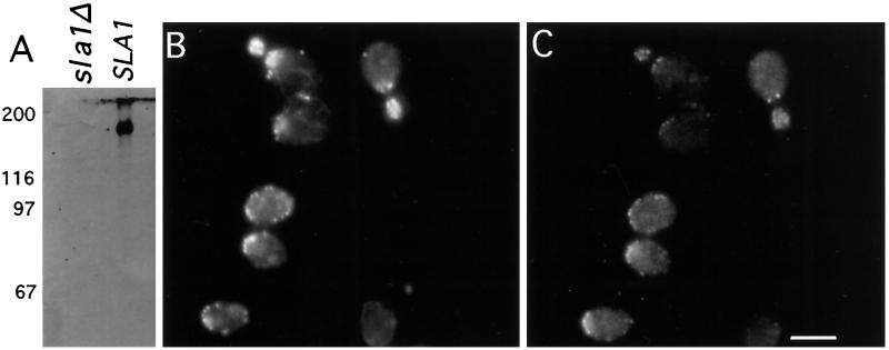
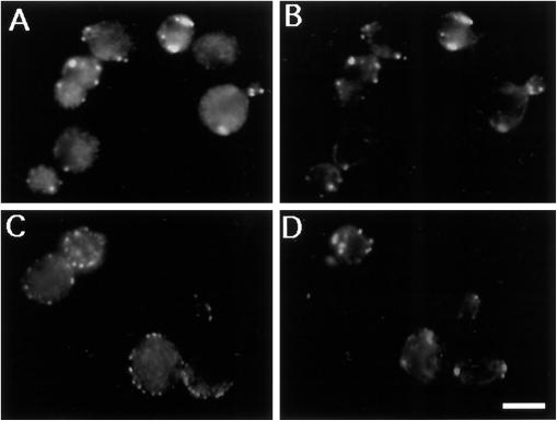
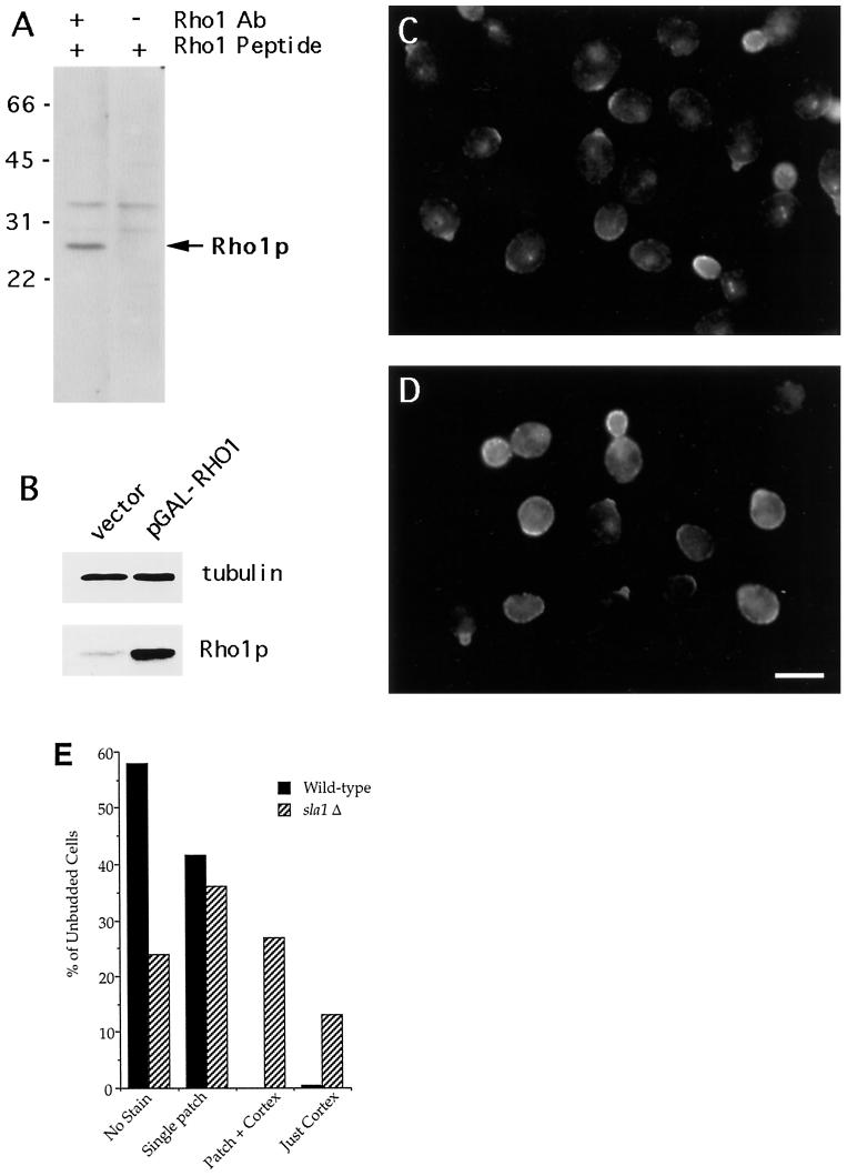
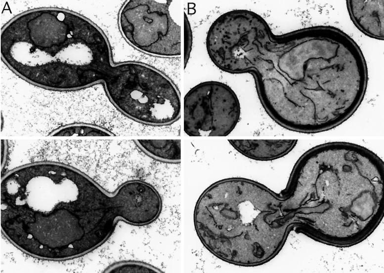
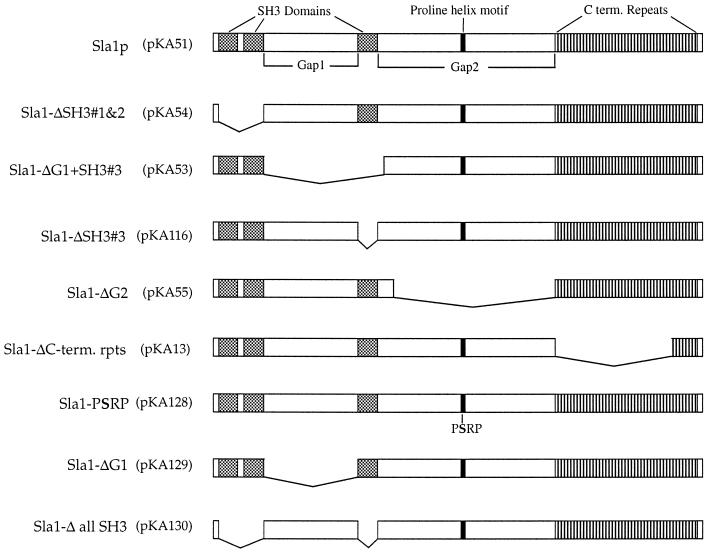
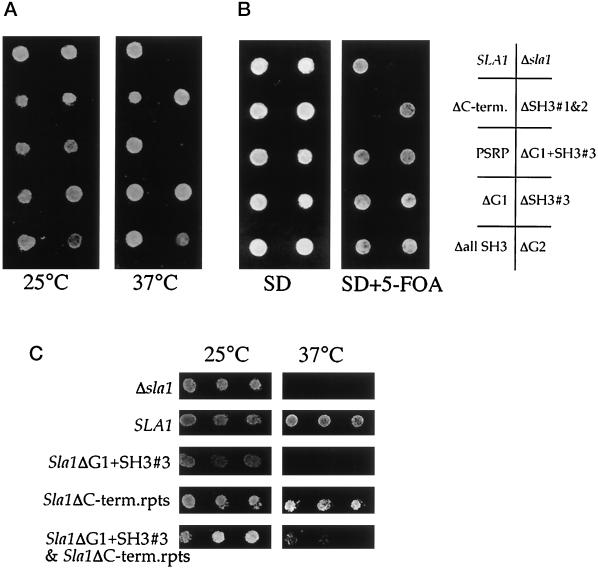
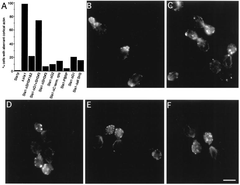
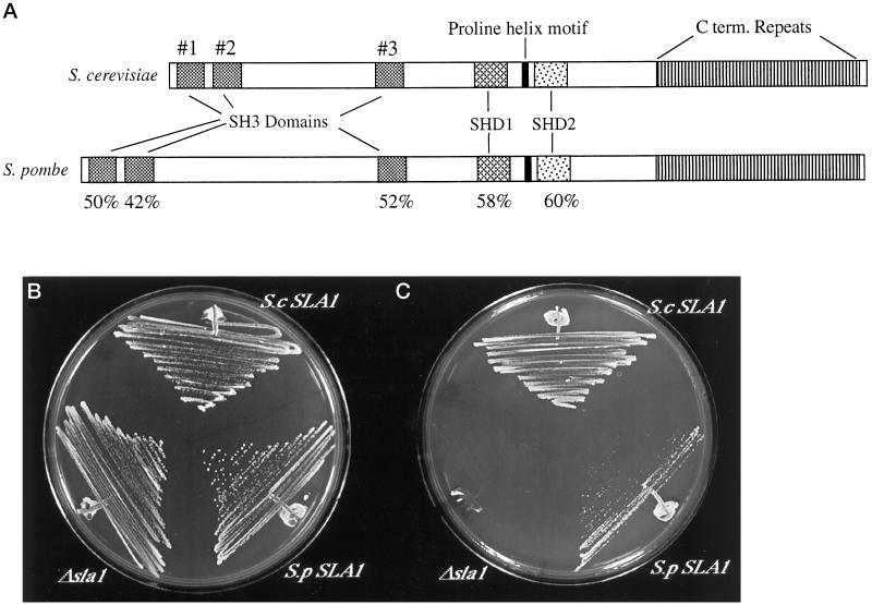
SLA1 was identified previously in budding yeast in a genetic screen for mutations that caused a requirement for the actin-binding protein Abp1p and was shown to be required for normal cortical actin patch structure and organization. Here, we show that Sla1p, like Abp1p, localizes to cortical actin patches. Furthermore, Sla1p is required for the correct localization of Sla2p, an actin-binding protein with homology to talin implicated in endocytosis, and the Rho1p-GTPase, which is associated with the cell wall biosynthesis enzyme beta-1,3-glucan synthase. Mislocalization of Rho1p in sla1 null cells is consistent with our observation that these cells possess aberrantly thick cell walls. Expression of mutant forms of Sla1p in which specific domains were deleted showed that the phenotypes associated with the full deletion are functionally separable. In particular, a region of Sla1p encompassing the third SH3 domain is important for growth at high temperatures, for the organization of cortical actin patches, and for nucleated actin assembly in a permeabilized yeast cell assay. The apparent redundancy between Sla1p and Abp1p resides in the C-terminal repeat region of Sla1p. A homologue of SLA1 was identified in Schizosaccharomyces pombe. Despite relatively low overall sequence homology, this gene was able to rescue the temperature sensitivity associated with a deletion of SLA1 in Saccharomyces cerevisiae.


Figures

Figure 1
Figure 1
Localization of Sla1p and actin using indirect double-label immunofluorescence microscopy. (A) Antibodies raised to Sla1p were used to detect Sla1p on Western blots of wild-type cells and to demonstrate lack of reactive proteins in sla1 null cells. (B and C) Immunofluorescence microscopy was then performed to localize Sla1p (C) and actin (B) in wild-type diploid cells. Bar, 5 μm.
Figure 2
Figure 2
Localization of proteins in the absence of Sla1p. sla1 null cells were grown to log phase and examined by double-label immunofluorescence microscopy. (A and B) Cells labeled for Abp1p (A) and actin (B). (C and D) Cells stained for Sla2p (C) and actin (D). Bar, 5 μm.
Figure 3
Figure 3
Deletion of SLA1 affects Rho1p localization. (A and B) Immunoblots of whole-cell lysates (10 μg/lane) probed with affinity-purified anti-Rho1p antibodies. (A) Lysate of wild-type cells (DDY664) probed with untreated antibodies (left lane) or with the same antibodies after preincubation with the Rho1p peptide antigen (right lane). (B) Lysates of DDY1097 (carrying a GALRHO1 plasmid; right lane) and DDY1098 (carrying a control plasmid) were prepared after growth on galactose for 8 h and probed with the anti-Rho1p antibodies (bottom) or (as a loading control) with anti-β-tubulin antibodies (top). (C and D) Immunolocalization of Rho1p in log-phase cells of wild-type (C) and sla1 null cells (D) Bar, 5 μm. (E) The staining patterns of unbudded cells were counted. The data shown are the averages of three experiments in which at least 200 unbudded cells were scored. The single patch of staining in wild-type unbudded cells is presumably the site from which the new bud will emerge.
Figure 4
Figure 4
sla1 null cells have thick cell walls. (A) Wild-type and (B) Δsla1 cells were grown to log phase and then fixed and processed for electron microscopy using potassium permanganate fixation to enhance visualization of the cell wall and membranes.
Figure 5
Figure 5
Mutant sla1 constructs. Details of plasmid construction are given in Table 2.
Figure 6
Figure 6
Growth of cells containing mutant Sla1 proteins. (A) Cells in which the genomic copy of SLA1 was deleted (KAY14) were transformed with the sla1 mutant constructs and plated onto selective medium at 25° and 37°C. (B) Cells carrying ABP1 on a URA3-marked plasmid (KAY78) were transformed with the sla1 mutant constructs and tested for growth on selective medium in the presence or absence of 5-FOA. (C) Cells (KAY14) were cotransformed with sla1 mutant constructs Sla1-ΔC-term.rpts and Sla1ΔGap1+SH3#3 and tested for growth at 37°C. Expression of both forms of Sla1p was verified by Western blotting.
Figure 7
Figure 7
Actin organization in cells expressing mutant forms of Sla1p. Log-phase cells were processed for Rd–phalloidin staining. (A) For each mutant at least 300 cells were scored for wild-type or aberrant actin patch morphology. Representative images of actin staining are shown for cells expressing (B) wild-type Sla1p, (C) no Sla1p, (D) Sla1-ΔG1+SH3#3, (E) Sla1-ΔC-terminal repeats, (F) Sla1-ΔSH3#1&2. Bar, 5 μm.
Figure 8
Figure 8
Rhodamine–actin incorporation into permeabilized cells expressing mutant forms of Sla1p. Cells expressing (A) wild-type Sla1p, (B) no Sla1p, (C) Sla1-ΔSH3#1&2, (D) Sla1-ΔG1+SH3#3, (E) Sla1-ΔC-term. repeats were grown, permeabilized, and tested for actin assembly as described by Li et al., (1995) (see MATERIALS AND METHODS). Sites of incorporation were then viewed by fluorescence microscopy, and the intensity of fluorescence in the buds was measured. The units of fluorescence intensity are arbitrary. Bar, 5 μm.
Figure 9
Figure 9
The S. pombe homologue of SLA1 rescues the temperature sensitivity of S. cerevisiae sla1Δ cells. (A) S. pombe Sla1p has a domain structure similar to that of S. cerevisiae Sla1p. Two regions of high homology are designated SHD (Sla1p homology domain) 1 and 2. The percentages of identity of the SH3 domains and the SHD regions are noted beneath the respective motifs. Growth of S. cerevisiae sla1 null cells (KAY14) containing S. cerevisiae SLA1, S. pombe sla1, or vector alone was tested at (B) 29°C and (C) 37°C.

References

    1. Ayscough KR. In vivo functions of actin-binding proteins. Curr Opin Cell Biol. 1998;10:102–111. - PubMed
    1. Ayscough KR, Drubin DG. Immunofluorescence microscopy of yeast cells. In: Celis J, editor. Cell Biology: A Laboratory Handbook. Vol. 2. San Diego: Academic Press; 1997. pp. 477–485.
    1. Ayscough KR, Stryker J, Pokala N, Sanders M, Crews P, Drubin DG. High rates of actin filament turnover in budding yeast and roles for actin in establishment and maintenance of cell polarity revealed using the actin inhibitor latrunculin-A. J Cell Biol. 1997;137:399–416. - PMC - PubMed
    1. Baudin A, Ozier-Kalogeropoulos O, Denoul A, Lacroute F, Cullin C. A simple and efficient method for direct gene deletion in Saccharomyces cerevisiae. Nucleic Acids Res. 1993;21:3329–3330. - PMC - PubMed
    1. Belmont LD, Drubin DG. The yeast V159N actin mutant reveals roles for actin dynamics in vivo. J Cell Biol. 1998;142:1289–1299. - PMC - PubMed

Publication types

MeSH terms