. 1999 Apr;10(4):1235-45.
doi: 10.1091/mbc.10.4.1235.

Architecture of the nuclear periphery of rat pachytene spermatocytes: distribution of nuclear envelope proteins in relation to synaptonemal complex attachment sites

Affiliations
Free PMC article

Architecture of the nuclear periphery of rat pachytene spermatocytes: distribution of nuclear envelope proteins in relation to synaptonemal complex attachment sites

M Alsheimer et al. Mol Biol Cell. 1999 Apr.
Free PMC article

Abstract

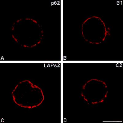
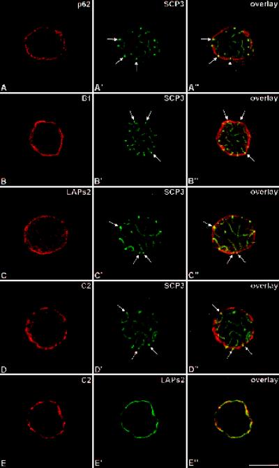
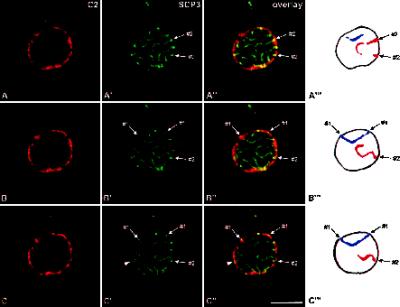
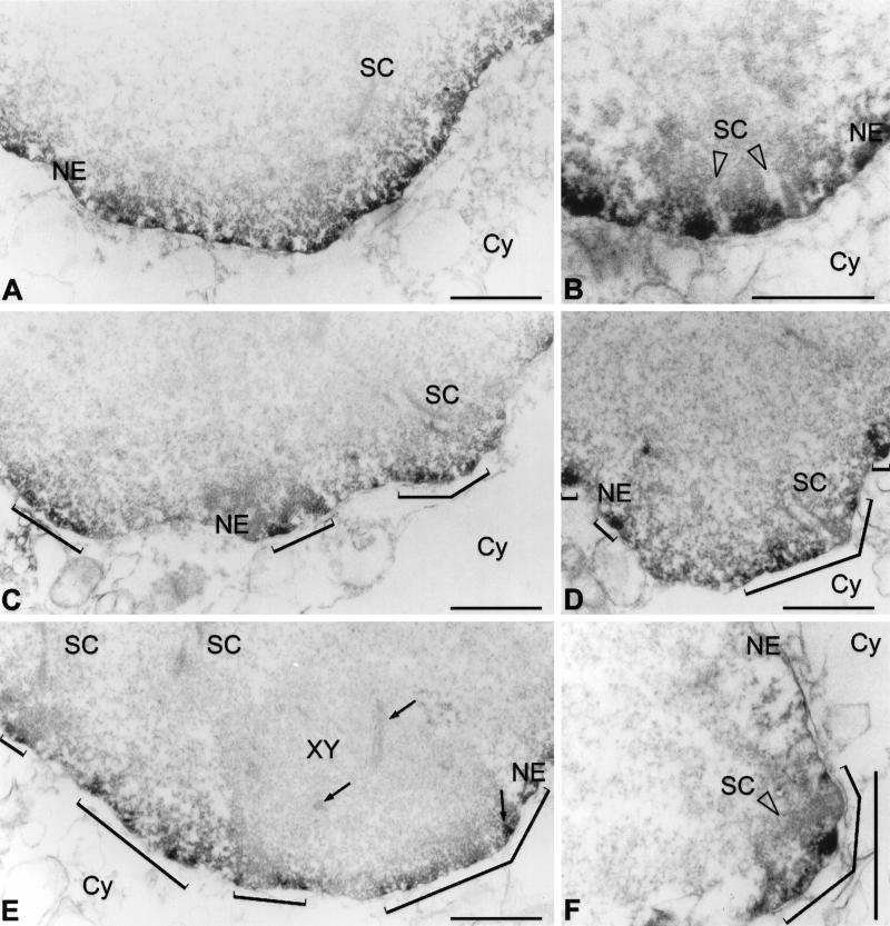
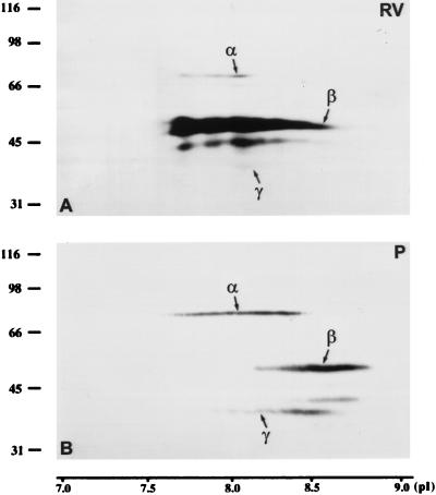
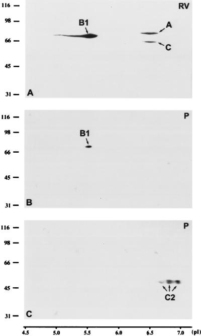
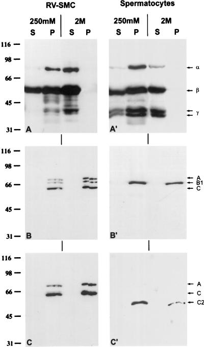
The nucleus of spermatocytes provides during the first meiotic prophase an interesting model for investigating relationships of the nuclear envelope (NE) with components of the nuclear interior. During the pachytene stage, meiotic chromosomes are synapsed via synaptonemal complexes (SCs) and attached through both ends to the nuclear periphery. This association is dynamic because chromosomes move during the process of synapsis and desynapsis that takes place during meiotic prophase. The NE of spermatocytes possesses some peculiarities (e.g., lower stability than in somatic cells, expression of short meiosis-specific lamin isoforms called C2 and B3) that could be critically involved in this process. For better understanding of the association of chromosomes with the nuclear periphery, in the present study we have investigated the distribution of NE proteins in relation to SC attachment sites. A major outcome was the finding that lamin C2 is distributed in the form of discontinuous domains at the NE of spermatocytes and that SC attachment sites are embedded in these domains. Lamin C2 appears to form part of larger structures as suggested by cell fractionation experiments. According to these results, we propose that the C2-containing domains represent local reinforcements of the NE that are involved in the proper attachment of SCs.


Figures

Figure 1
Figure 1
Immunolocalization of pore complex protein p62 (A), lamin B1 (B), LAPs2 (C) as well as lamin C2 (D). Labeled rat pachytene spermatocytes were investigated by confocal laser scanning microscopy. Bar, 10 μm.
Figure 2
Figure 2
Double-label immunolocalization with antibodies against pore complex protein p62 and SC-protein SCP3 (A–A′), lamin B1 and SCP3 (B–B′), LAPs2 and SCP3 (C–C′), lamin C2 and SCP3 (D–D′), and C2 and LAPs2 (E–E′). Labeled rat pachytene spermatocytes were investigated by confocal laser scanning microscopy. Overlays are shown in A"–E". Some of the attachment sites of the SCs are denoted by arrows. Bar, 10 μm.
Figure 3
Figure 3
Double-label immunolocalization with antibodies against lamin C2 (A–C) and protein SCP3 (A′–C′). The labeled rat pachytene spermatocyte was investigated by confocal laser scanning microscopy. Three consecutive optical sections are shown (A–A′, B–B′, C–C′). Overlays are presented in A"–C". A‴–C‴, Schematic representation of the arcs displayed by SCs #1 (blue) and #2 (red). The arrowheads in C′–C" point at an XY body. Bar, 10 μm.
Figure 4
Figure 4
Electron microscopical immunolocalization of lamins B1 (A, B) and C2 (C–F) in pachytene spermatocytes of the rat using peroxidase-conjugated secondary antibodies. The patchy pattern obtained with C2 antibodies is denoted by brackets (C–F). Frontal (B, D) and lateral (F) views of SC attachment sites at the NE are shown. XY, XY body; Cy, cytoplasm. XY body axial elements (including an insertion at the NE) are denoted by arrows (E). Bars, 1 μm.
Figure 5
Figure 5
Two-dimensional PAGE of whole RV-SMC cells (A; 5 × 10⁶ cells) and pachytene spermatocytes of the rat (B; 5 × 10⁶ cells). After transfer to nitrocellulose, the proteins were incubated with mAb 13d4 against the LAPs2 α, β, and γ.
Figure 6
Figure 6
Two-dimensional PAGE of whole RV-SMC cells (A; 5 × 10⁶ cells) and pachytene spermatocytes of the rat (B, C; 5 × 10⁶ cell/gel). After transfer to nitrocellulose, the proteins were incubated with mAb PKB8 against lamins A, B1, and C (A and B) or mAb R27 against lamins A, C, and C2 (C).
Figure 7
Figure 7
Immunoblotting of NE proteins of RV-SMC cells (A–C; 2.5 × 10⁴ cell equivalents/lane) and pachytene spermatocytes of the rat (A′–C′; 2.5 × 10⁴ cell equivalents/lane) that were fractionated with buffers containing 1% Triton X-100 and 250 mM or 2 M salt. mAbs 13d4 (A–A′; against LAPs2 α, β, and γ), PKB8 (B–B′; against lamins A, B1, and C) as well as R27 (C–C′; against lamins A, C, and C2) were used to detect the respective antigens. S, Supernatant fraction; P, pellet fraction.

References

    1. Alsheimer M, Benavente R. Change of karyoskeleton during mammalian spermatogenesis: expression pattern of nuclear lamin C2 and its regulation. Exp Cell Res. 1996;228:181–188. - PubMed
    1. Alsheimer M, Fecher E, Benavente R. Nuclear envelope remodelling during rat spermiogenesis: distribution and expression pattern of LAP2/thymopoietins. J Cell Sci. 1998;111:2227–2234. - PubMed
    1. Bass HW, Marshall WF, Sedat JW, Agard DA, Cande WZ. Telomeres cluster de novo before the initiation of synapsis: a three-dimensional spatial analysis of telomere positions before and during meiotic prophase. J Cell Biol. 1997;137:5–18. - PMC - PubMed
    1. Belmont AS, Zhai Y, Thilenius A. Lamin B distribution and association with peripheral chromatin revealed by optical sectioning and electron microscopic tomography. J Cell Biol. 1993;123:1671–1685. - PMC - PubMed
    1. Benavente R, Dabauvalle MC, Scheer U, Chaly N. Functional role of newly formed pore complexes in postmitotic nuclear reorganization. Chromosoma. 1989;98:233–241. - PubMed

Publication types

LinkOut - more resources