. 1999 Apr 13;96(8):4313-8.
doi: 10.1073/pnas.96.8.4313.

A MHC-encoded ubiquitin-like protein (FAT10) binds noncovalently to the spindle assembly checkpoint protein MAD2

Affiliations

A MHC-encoded ubiquitin-like protein (FAT10) binds noncovalently to the spindle assembly checkpoint protein MAD2

Y C Liu et al. Proc Natl Acad Sci U S A. .

Abstract

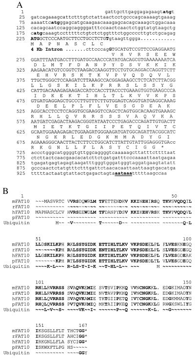
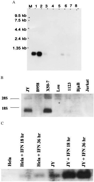
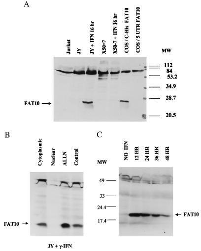
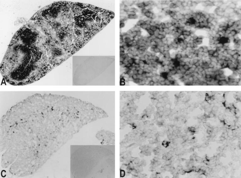
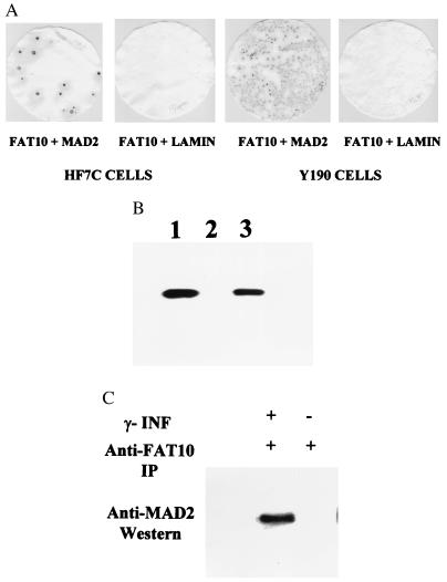
Recently a number of nonclass I genes were discovered in the human MHC class I region. One of these, FAT10, encodes a protein consisting of two domains with homology to ubiquitin. FAT10 mRNA is expressed constitutively in some lymphoblastoid lines and dendritic cells and in certain other cells after gamma-interferon induction. FAT10 protein expression is controlled at several levels including transcription, translation, and protein stability. Yeast two-hybrid screening of a human lymphocyte library and immunoprecipitation studies revealed that FAT10 noncovalently associated with MAD2, a protein implicated in a cell-cycle checkpoint for spindle assembly during anaphase. Thus, FAT10 may modulate cell growth during B cell or dendritic cell development and activation.


Figures

Figure 1
Figure 1
The sequence of human FAT10 gene. (A) The human FAT10 gene contains 971 base pairs spanning two exons. There are three short uORFs (starting at nucleotides 21, 82, and 176) preceding the long FAT10 ORF (225 to 722). Potential translation initiation codons for the short uORF are indicated by lower-case bold letters and that for FAT10 by upper-case bold letters. The FAT10 coding sequence represented by capital letters encodes a 166-aa polypeptide. The four-kilobase intron is located between codons 9 and 10, represented by bold letters. The poly(A) signal of AATAAA is underlined at the 3′ end of FAT10 gene. The access number for this sequence is AF123050. (B) Comparison of human, murine, partial rat, and porcine FAT10 proteins with classical ubiquitin amino acids sequences. The ubiquitin sequence is shown twice to compare with the two FAT10 UBL domains, and only the conserved amino acids are presented in the diagram.
Figure 2
Figure 2
Northern blot analysis of FAT10 mRNA. (A) The multiple human normal tissues Northern blot (CLONTECH). Lane 1, spleen; 2, thymus; 3, prostate; 4, testis; 5, ovary; 6, small intestine; 7, colon mucosas lining, and 8, peripheral blood leukocytes. Lane M is the RNA marker as labeled. (B) The analysis of total RNA blots from various cell lines. The RNA preparation and hybridization were performed as described (29). The origins of cell lines are indicated at the top and the two ribosomal RNA bands are indicated at the side as 28S and 18S. (C) γ-IFN treatment (3,000 u/ml) (0, 18, and 36 hr) induces progressive increases in FAT10 mRNA expression in JY and HeLa cells.
Figure 3
Figure 3
Western analysis of FAT10 proteins in various cell lines. (A) The Western analysis of JY and X50–7 cells in 20% SDS/PAGE shows that γ-IFN (3,000 u/ml) can induce FAT10 protein in JY cells, but not in Jurkat or γ-IFN-treated X50–7 cells. The C-His-FAT10 construct produces a single his-tagged FAT10 protein in transfected COS cells, while the uORF-FAT10 produces no detectable FAT10 protein. (B) Western analysis of FAT10 protein in 12% SDS/PAGE shows that the majority of FAT10 protein induced by γ-IFN is in the cytoplasmic fraction. Short treatment (1.5 hr) of JY cells with ALLN (260 μM) after overnight γ-IFN induction also increases the FAT10 protein level. (C) Induction of FAT10 protein by γ-IFN with JY cells peaks between 12 and 24 hr induction and then gradually declines with time.
Figure 4
Figure 4
In situ hybridization on mouse tissue sections. Mouse thin sections (5 μ) were hybridized with FAT10 antisense (A and B, spleen; C and D, thymus) and sense probes (insets in A and C). Expression of murine FAT10 mRNA was seen only with antisense probe. The heavily stained cells are scattered in the white pulp in the spleen (A and B). In mouse thymus, the positive cells were mainly in cortico-medullary junction (C). At higher resolution (D), most FAT10 mRNA was in cells with expanded cytoplasm.
Figure 5
Figure 5
The specific interaction of FAT10 with MAD2 in a yeast two-hybrid screen (A), in vitro on a GST column (B), and in vivo in immunoprecipitates (C). (A) The β-galactosidase assays show positive (blue) for specific interaction between FAT10 and MAD2 but negative (white) for FAT10 and human lamin C. (B) The FAT10-GST fusion proteins were produced with the GST expression vector, pGEX-4T-1 (Pharmacia). The 35S-labeled MAD2 was added to the GST column in RPMI 1640 medium. The 35S-labeled MAD2 retained on GST columns was washed, eluted, and analyzed in 10% SDS/PAGE. (1) 35S- labeled MAD2 control, (2) 35S MAD2 eluted from GST protein bound GST column, (3) 35S MAD2 from FAT10-GST bound GST column. (C) Anti-FAT10 polyclonal antibody was used to immunoprecipitate FAT10 in the JY cell lysates in 50 mM Tris (pH 8)/150 mM NaCl/1% Triton X-100/100 μg/ml PMSF and a protease inhibitor mixture (Boehringer Mannheim). The antibody–FAT10 complex was pulled down with protein A Sepharose 6B (Pharmacia) resin. After extensive wash, the final immunoprecipitate was analyzed by Western blot with anti-MAD2 antibody (Santa Cruz Biotechnology). The MAD2 clearly coprecipitates with FAT10 in the γ-IFN induced JY cell lysate but not in the uninduced JY cell lysate.

Similar articles

Cited by

References

    1. Gruen J R, Weissman S M. Blood. 1997;90:4252–4265. - PubMed
    1. Gruen J R, Nalabolu S R, Chu T W, Bowlus C, Fan W, Goei V L, Wei H, Sivakamasundari R, Liu Y-C, Xu H, et al. Genomics. 1996;36:70–85. - PubMed
    1. Fan W, Cai W, Parimoo S, Lennon G G, Weissman S M. Immunogenetics. 1996;44:97–103. - PubMed
    1. Bates E E, Ravel O, Dieu M C, Ho S, Guret C, Bridon J M, Ait-Yahia S, Briere F, Caux C, Banchereau J, et al. Eur J Immunol. 1977;10:2471–2477. - PubMed
    1. Hochstrasser M. Annu Rev Genet. 1997;30:405–439. - PubMed

Publication types

MeSH terms

Associated data