Comparative Study
. 1999 Apr 13;96(8):4319-24.
doi: 10.1073/pnas.96.8.4319.

Crystal structure of human T cell leukemia virus type 1 gp21 ectodomain crystallized as a maltose-binding protein chimera reveals structural evolution of retroviral transmembrane proteins

Affiliations
Comparative Study

Crystal structure of human T cell leukemia virus type 1 gp21 ectodomain crystallized as a maltose-binding protein chimera reveals structural evolution of retroviral transmembrane proteins

B Kobe et al. Proc Natl Acad Sci U S A. .

Abstract

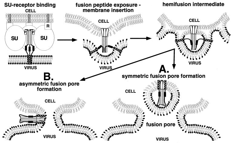
Retroviral entry into cells depends on envelope glycoproteins, whereby receptor binding to the surface-exposed subunit triggers membrane fusion by the transmembrane protein (TM) subunit. We determined the crystal structure at 2.5-A resolution of the ectodomain of gp21, the TM from human T cell leukemia virus type 1. The gp21 fragment was crystallized as a maltose-binding protein chimera, and the maltose-binding protein domain was used to solve the initial phases by the method of molecular replacement. The structure of gp21 comprises an N-terminal trimeric coiled coil, an adjacent disulfide-bonded loop that stabilizes a chain reversal, and a C-terminal sequence structurally distinct from HIV type 1/simian immunodeficiency virus gp41 that packs against the coil in an extended antiparallel fashion. Comparison of the gp21 structure with the structures of other retroviral TMs contrasts the conserved nature of the coiled coil-forming region and adjacent disulfide-bonded loop with the variable nature of the C-terminal ectodomain segment. The structure points to these features having evolved to enable the dual roles of retroviral TMs: conserved fusion function and an ability to anchor diverse surface-exposed subunit structures to the virion envelope and infected cell surface. The structure of gp21 implies that the N-terminal fusion peptide is in close proximity to the C-terminal transmembrane domain and likely represents a postfusion conformation.


Figures

Figure 1
Figure 1
(A) Structure determination. Electron density map (22) (magenta) calculated with coefficients |Fobs| − |Fcalc| (40–2.5 Å resolution) and phases based on the MBP positioned by molecular replacement, contoured at 1.5 SDs. Superimposed is the Cα trace of the refined model of MBP/gp21 trimer (MBP green, gp21 yellow). The path of the gp21 chain is clearly visible in this map. (B) Structure of MBP/gp21 chimera. Ribbon diagram of a trimer [drawn with programs molscript (48) and raster3d (49)]. MBP is green, the three-alanine linker is yellow, and gp21 is blue. The view is down the crystallographic 3-fold axis (rotated 90° around the horizontal axis relative to the view in A).
Figure 2
Figure 2
Structure of gp21. (a) Ribbon diagram of the gp21 monomer with the coiled coil-forming helix magenta, the disulfide-bonded domain CX6CC green, the C-terminal helix cyan, and the rest gray. The side chains of Met-338, Gly-343, Leu-346, and Val-350 with unusual packing (yellow), the a and d residues in the coiled coil (magenta), the hydrophobic residues in the base of the coiled coil (gray), and the cysteine residues (green) are shown. (b) As in a but showing trimer. (c) Ribbon diagram of MoMLV TM trimer (8) with the coiled coil-forming helix magenta and the disulfide-bonded domain CX6CC green. The side chains of the a and d residues in the coiled coil (magenta), the hydrophobic residues in the base of the coiled coil (gray), and the cysteine residues (green) are shown. (d) Ribbon diagram of HIV-1 gp41 trimer (10) with the coiled coil-forming helix magenta and the C-terminal helix cyan. The side chains of Gln-41 in the glutamine-rich layer (yellow), and the a and d residues in the coiled coil (magenta) are shown. GCN4 region is not shown. (e) Ribbon diagram of the TBHA2 trimer (26) with the coiled coil-forming helix magenta, the C-terminal helix cyan, the HA1 residues blue, and the rest gray. The side chains of Thr-59 with x-type packing in a region where loop-to-helix transition occurs at low pH (yellow), and the a and d residues in the coiled coil (magenta) are shown. (f) Molecular surface (probe radius 1.4 Å) of the trimeric coiled coil of gp21 (residues 338–387) color-coded according to surface complementarity (50) with the C-terminal segment (residues 390–421). Red, Sc (shape correlation statistic) > 0.76; yellow, 0.76 > Sc > 0.3; green, 0.3 > Sc > −0.3; light blue, Sc < −0.3. The overall Sc equals 0.65. The C-terminal segment is shown in magenta, with the interacting residues (contacts < 4 Å) in white. Drawn by grasp (51).
Figure 3
Figure 3
Structural diversity of retroviral TMs. Comparison of known (cylinders; gp21, red; MoMLV TM, light green; gp41, dark green) and predicted (boxed) (31) α-helices present in the TM ectodomains of the retroviridae. Extended/random coil is shown as a green line and 310-helix as a blue cylinder. Residues in the a and d positions of the heptad repeat are highlighted in magenta and residues involved in irregular packing at the N terminus of the coiled coil are highlighted in yellow. Acidic residues are red, basic residues blue, aromatic residues cyan, and cysteines highlighted in olive. Known and potential N-linked glycosylation sites are italic and underlined. The retrovirus type species used are HTLV-13–19-3 (HTLV), STLV-L (STLV, GenBank accession no. Y07616), BLV (K02120), MoMLV (J02255), SMRV, (M23385); MPMV (AF033815), RSV (AF033808); HIV-1HXB2R (HIV, K03455), SIVAGMtan (SIV, U58991); and mouse mammary tumor virus (MMTV) (D16249). *RSV has an internal fusion peptide: IFASILAPGVAAA.
Figure 4
Figure 4
Model of retroviral membrane fusion in which the trimeric symmetry of the TM is maintained during the fusion process. R, cellular receptor. In the TM subunit, fusion peptides are indicated by white cylinders marked F, the N-terminal coiled coil by light gray cylinders, the C-terminal segment is black and the transmembrane domain is hatched. The cytoplasmic tails are not shown. Two scenarios (A and B) are possible for fusion pore formation (see text). The model is based on evidence accumulated for membrane fusion in retroviruses and influenza virus; experimental evidence for conformational changes in gp21 induced by receptor binding is not yet available.

Similar articles

Cited by

References

    1. Poiesz B J, Ruscetti F W, Gazdar A F, Bunn P A, Minna J D, Gallo R C. Proc Natl Acad Sci USA. 1980;77:7415–7419. - PMC - PubMed
    1. Osame M, Usuku K, Izumo S, Ijishi N, Amitani H, Igata A, Matsumoto M, Tara M. Lancet. 1986;1:1031–1032. - PubMed
    1. Fan N, Gavalchin J, Paul B, Wells K, Lane M J, Poiesz B. J Clin Microbiol. 1992;30:905–910. - PMC - PubMed
    1. Donegan E, Lee H, Operskalski E A, Shaw G M, Kleinman S H, Busch M P, Stevens C E, Schiff E R, Nowicki M J, Hollingsworth C G, et al. Transfusion. 1994;34:478–483. - PubMed
    1. Pique C, Pham D, Tursz T, Dokhélar M-C. J Virol. 1992;66:906–913. - PMC - PubMed

Publication types

MeSH terms

Substances

Associated data

LinkOut - more resources