. 1999 Apr 13;96(8):4432-7.
doi: 10.1073/pnas.96.8.4432.

Tamoxifen inhibits acidification in cells independent of the estrogen receptor

Affiliations

Tamoxifen inhibits acidification in cells independent of the estrogen receptor

N Altan et al. Proc Natl Acad Sci U S A. .

Abstract

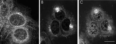
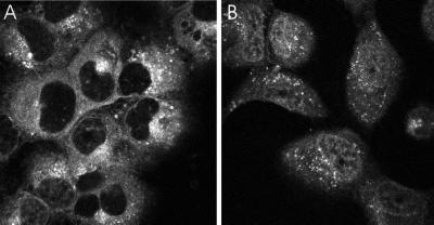
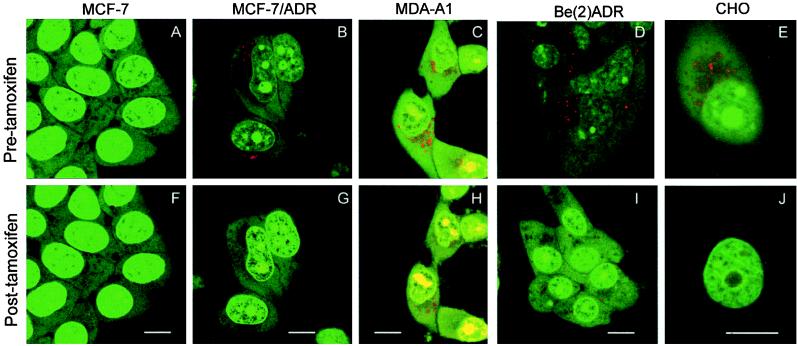
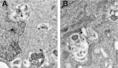
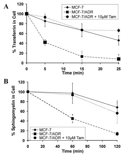
Tamoxifen has been reported to have numerous physiological effects that are independent of the estrogen receptor, including sensitization of resistant tumor cells to many chemotherapeutic agents. Drug-resistant cells sequester weak base chemotherapeutics in acidic organelles away from their sites of action in the cytosol and nucleus. This work reports that tamoxifen causes redistribution of weak base chemotherapeutics from acidic organelles to the nucleus in drug-resistant cells. Agents that disrupt organelle acidification (e.g., monensin, bafilomycin A1) cause a similar redistribution. Measurement of cellular pH in several cell lines reveals that tamoxifen inhibits acidification of endosomes and lysosomes without affecting cytoplasmic pH. Similar to monensin, tamoxifen decreased the rate of vesicular transport though the recycling and secretory pathways. Organellar acidification is required for many cellular functions, and its disruption could account for many of the side effects of tamoxifen.


Figures

Figure 1
Figure 1
Steady-state distribution of Adriamycin (A) in drug-sensitive MCF-7 cells was diffuse throughout the cytoplasm with an increased fluorescence in the nucleoplasm. (B) In drug-resistant MCF-7/ADR cells Adriamycin is excluded from the nucleus and is accumulated in punctate cytoplasmic organelles. (C) The MCF-7/ADR cells in B were treated with 10 μM tamoxifen for 30 min, resulting in a redistribution of Adriamycin from the cytoplasmic organelles to the nucleus.
Figure 2
Figure 2
Washout of Adriamycin from MCF-7/ADR cells. MCF-7/ADR cells were incubated with 10 μM Adriamycin for 30 min and then rinsed free of external Adriamycin in the absence (A) or presence (B) of tamoxifen. (A) In the absence of tamoxifen, Adriamycin fluorescence slowly decreased after washout. (B) The presence of tamoxifen caused decrease in punctate fluorescence in increase in nuclear fluorescence, indicating that Adriamycin diffused out of the punctate compartments into the nucleus and cytoplasm.
Figure 3
Figure 3
AO labeling of MCF-7/ADR cells. AO accumulates in acidic compartments, producing a red fluorescence. (A) In drug-sensitive MCF-7 cells the AO fluorescence was a relatively even green with no red-orange fluorescence. Punctate red-orange fluorescence, indicative of acidified organelles, was observed in the cytoplasm of: (B) drug-resistant MCF-7/ADR breast tumor line, (C) drug-resistant (estrogen-receptor negative) MDA-A1 cells breast tumor line, (D) drug-resistant Be(2)ADR neuroblastoma cell line, and (E) Chinese hamster ovary (CHO) cell line. (F) Tamoxifen had no effect on the fluorescence of MCF-7 cells. However, 30 min after the addition of 5 μM tamoxifen there was a reduction in the red-orange fluorescence in the (G) MCF-7/ADR cells, (H) MDA-A1 cells, (I) Be(2)ADR cells, and (J) CHO cells. Cells were incubated with 2 μM AO as described in Materials and Methods and examined under laser-scanning confocal microscopy. (Bar is 10 μm.)
Figure 4
Figure 4
Effect of tamoxifen on DAMP labeling of MCF-7/ADR cells. Acidification of the lysosomes was probed with the weak base DAMP, which accumulates in acidic organelles. (A) Electron micrograph of mouse anti-DNP and gold-conjugated anti-mouse antibodies in MCF-7/ADR cells. The gold particles indicate accumulation of DAMP within cytoplasmic organelles. The average density of gold particles was 7.02/μm2 of lysosomal area. (B) Cells incubated with tamoxifen had a substantial reduction of anti-DAMP labeling to 2.0 gold particles/μm2 of lysosomal area. Cells were incubated with DAMP, then fixed and prepared for immunoelectron microscopy as described in Materials and Methods. (Bar is 1 μm.)
Figure 5
Figure 5
Effect of tamoxifen on transport in MCF-7/ADR cells. (A) Kinetics of transport of BODIPY-transferrin to the surface. The cell-associated BODIPY-transferrin was quantified at various time points by using confocal microscopy. After 5 min only 50% of the transferrin was still associated with the MCF-7/ADR cells (■). In contrast, more than 90% remained with MCF-7/ADR cells that had been incubated with 10 μM tamoxifen (●). After 25 min less than 10% of the transferrin remained with the control MCF-7/ADR cells and more than 60% remained with the tamoxifen-treated cells. The rate of transferrin transport in MCF-7/ADR cells treated with tamoxifen was similar to the rate in drug-sensitive MCF-7 cells (⧫). (B) Kinetics of transport of BODIPY-sphingomyelin to the surface. The kinetics of transport of the lipid sphingomyelin from the TGN to the surface was quantified as described in Materials and Methods. Two hours after removal of the BODIPY-ceramide, the fluorescence in the MCF-7/ADR cells decreased to almost 20% (■). In the presence of tamoxifen (10 μM) the BODIPY-fluorescence decrease was slower (●). In the MCF-7 cells (⧫) the rate of transport of the BODIPY-sphingomyelin to the surface was similar to that of the MCF-7/ADR cells with tamoxifen.

Similar articles

Cited by

References

    1. Spiegel D. Semin Oncol. 1997;24:S1-36–S1-47. - PubMed
    1. Jaiyesimi I A, Buzdar A U, Decker D A, Hortobagyi G N. J Clin Oncol. 1995;13:513–529. - PubMed
    1. Early Breast Cancer Trialists’ Collaborative Group. Lancet. 1998;351:1451–1467. - PubMed
    1. Jordan V C. Proc Soc Exp Biol Med. 1995;208:144–149. - PubMed
    1. Fisher B, Costantino J P, Wickerham D L, Redmond C K, Kavanah M, Cronin W M, Vogel V, Robidoux A, Dimitrov N, Atkins J, et al. J Natl Cancer Inst. 1998;90:1371–1388. - PubMed

Publication types

MeSH terms