. 1999 Apr 13;96(8):4546-51.
doi: 10.1073/pnas.96.8.4546.

Distribution of human herpesvirus-8 latently infected cells in Kaposi's sarcoma, multicentric Castleman's disease, and primary effusion lymphoma

Affiliations

Distribution of human herpesvirus-8 latently infected cells in Kaposi's sarcoma, multicentric Castleman's disease, and primary effusion lymphoma

N Dupin et al. Proc Natl Acad Sci U S A. .

Abstract

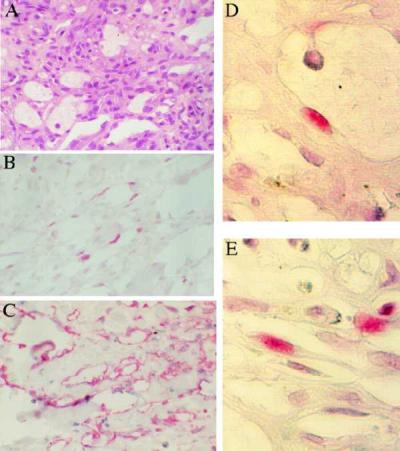
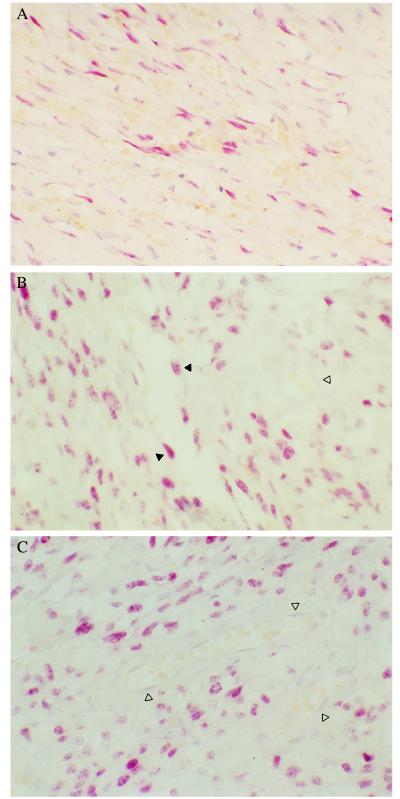
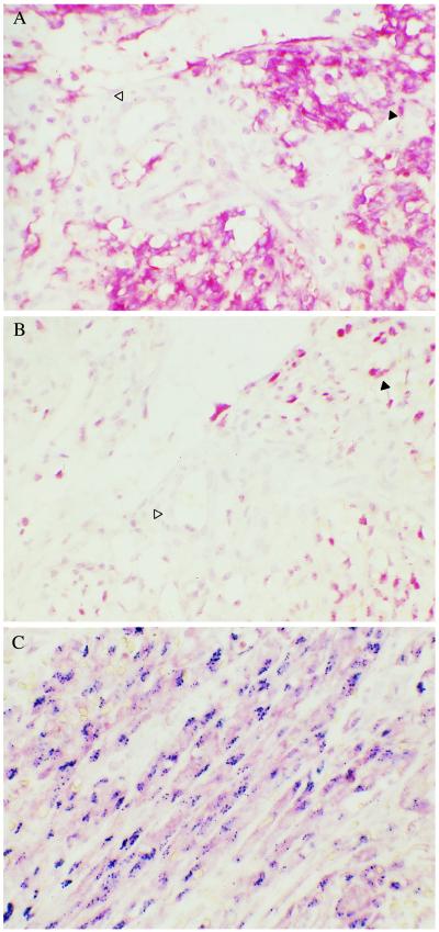
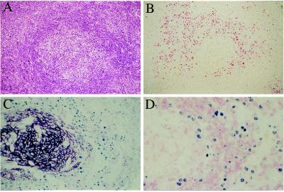
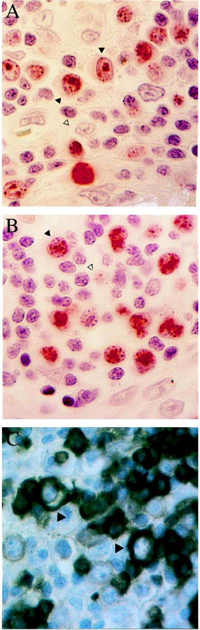
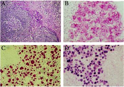
Human herpesvirus 8 (HHV-8, also called KSHV) is linked to the etiopathogenesis of Kaposi's sarcoma (KS), multicentric Castleman's disease (MCD), and primary effusion lymphoma (PEL). The universal presence of HHV-8 in early KS has not yet been shown. We used a mAb (LN53) against latent nuclear antigen-1 (LNA-1) of HHV-8 encoded by ORF73 to study the distribution of the cell types latently infected by HHV-8 in patch, plaque, and nodular KS, MCD, and PEL. In early KS, HHV-8 is present in <10% of cells forming the walls of ectatic vessels. In nodular KS, HHV-8 is present in cells surrounding slit-like vessels and in >90% of spindle cells, but not in normal vascular endothelium. In addition, HHV-8 colocalizes with vascular endothelial growth factor receptor-3 (VEGFR-3), a marker of lymphatic and precursor endothelium. In early KS lesions, VEGFR-3 is more extensively expressed than LNA-1, indicating that HHV-8 is not inducing the proliferation of VEGFR-3-positive endothelium directly. In MCD, HHV-8 is present in mantle zone large immunoblastic B cells. No staining for LNA-1 is seen in samples from multiple myeloma, prostate cancer, and angiosarcoma, supporting the absence of any etiological link between these diseases and HHV-8.


Figures

Figure 1
Figure 1
Patch stage KS (×40). (A) Hematoxylin-eosin (H/E) stain of patch KS lesion. (B) LNA-1 staining of adjacent sections shows that latent HHV-8 infection is present in fusiform cells forming the walls of ectatic spaces; staining is nuclear. (C) VEGFR-3 staining of adjacent section shows expression by all cells forming the walls of ectatic vessels; staining is cytoplasmic. (D and E) Higher magnification (×160) of patch KS stained with LNA-1 showing (D) one positive cell in the wall of an ectatic vessel and (E) positive cells forming the walls of slit-like vessels.
Figure 2
Figure 2
(A) In a plaque KS lesion, up to 50% of spindle-shaped cells surrounding the typical lacs vasculaires are positive for HHV-8 by LN53 (×40). (B and C) Nodular KS stained for HHV-8 with LN53 (×40). >90% of spindle cells are positive. The typical nuclear stippling pattern is seen. Endothelial cells surrounding an angulated vessel are positive (black arrowheads). However, in both figures the endothelial cells surrounding mature vascular spaces are negative for HHV-8 (white arrowheads).
Figure 3
Figure 3
Expression of VEGFR-3 in nodular KS (×40). (A) Most spindle cells express VEGFR-3, and endothelial cells surrounding ectatic vessels are positive for VEGFR-3 (black arrowheads), whereas endothelial cells surrounding a normal blood vessel are negative (white arrowhead). (B) Adjacent section of A stained with LN53 shows that LNA-1 positive cells have a similar distribution to VEGFR-3, although fewer cells are positive for LNA-1. The same vessel negative for VEGFR-3 is also negative for LNA-1 (white arrowhead). (C) Costaining of VEGFR-3 (cytoplasmic, pink) and HHV-8 (nuclear, dark blue stippling) confirms that cells containing HHV-8 express VEGFR-3.
Figure 4
Figure 4
Expression of CD23, CD79a-cy, and HHV-8 LNA-1 in a lymph node biopsy from MCD. (A) H/E staining of a follicle (×10). (B) Adjacent section stained with LN53 showing many positive cells in the mantle zone (nuclear red) (×10). (C) Costaining with CD23 (cytoplasmic, dark purple) and LN53 (nuclear, light blue) confirms that the positive cells for HHV-8 do not express CD23 (×20). (D) Costaining with CD79a-cy (plasma membrane, pink) and LN53 (nuclear, blue) shows that HHV-8 positive cells do not express CD79 (×40).
Figure 5
Figure 5
High magnification (×140) of MCD mantle zone. (A and B) The HHV-8-positive cells (black arrowheads) are large, have prominent nuclei with one or two nucleoli, and resemble immuno- or plasmablastic cells. Staining for LNA-1 shows the typical nuclear stippling (black arrowhead). Lymphocytes surrounding the HHV-8-positive cells are negative (white arrowheads). (C) CD20 staining shows that the large immunoblastic cells are positive (black arrowhead).
Figure 6
Figure 6
(A) The lymph node of a patient with PEL stained with H/E showing large anaplastic cells (×20). (B and C) These neoplastic cells express CD30 (B) and HHV-8 LNA-1 (C) (×40). (D) Costaining confirms that CD30 (plasma membrane, red) and LNA-1 (nuclear stippling, purple) colocalize in the neoplastic cells (×40).

References

    1. Moore P S, Chang Y. Am J Epidemiol. 1998;147:217–221. - PubMed
    1. Boshoff C, Weiss R A. In: Advances in Cancer Research. Van de Woude G, Klein G, editors. Vol. 75. San Diego: Academic; 1998. pp. 57–86. - PubMed
    1. Boshoff C, Schulz T F, Kennedy M M, Graham A K, Fisher C, Thomas A, McGee J O, Weiss R A, O’Leary J J. Nat Med. 1995;1:1274–1278. - PubMed
    1. Li J J, Huang Y Q, Cockerell C J, Friedman Kien A E. Am J Pathol. 1996;148:1741–1748. - PMC - PubMed
    1. Foreman K E, Bacon P E, Hsi E D, Nickoloff B J. J Clin Invest. 1997;99:2971–2978. - PMC - PubMed

Publication types

MeSH terms

Substances