. 1999 Apr 13;96(8):4552-7.
doi: 10.1073/pnas.96.8.4552.

Defective CD95/APO-1/Fas signal complex formation in the human autoimmune lymphoproliferative syndrome, type Ia

Affiliations

Defective CD95/APO-1/Fas signal complex formation in the human autoimmune lymphoproliferative syndrome, type Ia

D A Martin et al. Proc Natl Acad Sci U S A. .

Erratum in

  • Proc Natl Acad Sci U S A. 2004 May 18;101(20):7840

Abstract

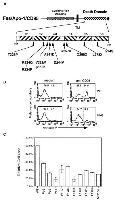
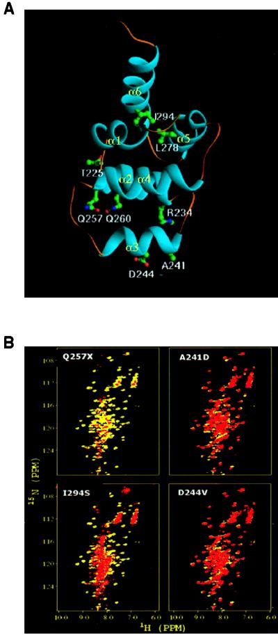
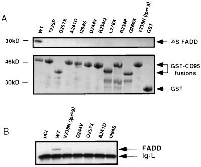
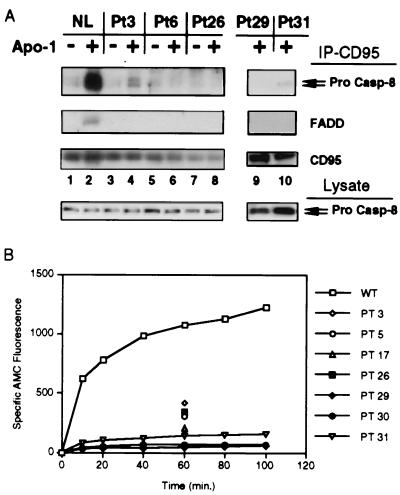
Heterozygous mutations in the CD95 (APO-1/Fas) receptor occur in most individuals with autoimmune lymphoproliferative syndrome (ALPS) and dominantly interfere with apoptosis by an unknown mechanism. We show that local or global alterations in the structure of the cytoplasmic death domain from nine independent ALPS CD95 death-domain mutations result in a failure to bind the FADD/MORT1 signaling protein. Despite heterozygosity for the abnormal allele, lymphocytes from ALPS patients showed markedly decreased FADD association and a loss of caspase recruitment and activation after CD95 crosslinking. These data suggest that intracytoplasmic CD95 mutations in ALPS impair apoptosis chiefly by disrupting death-domain interactions with the signaling protein FADD/MORT1.


Figures

Figure 1
Figure 1
CD95 “death-domain” amino acid changes in ALPS patients. (A) Diagram of the CD95 protein. The extracellular cysteine-rich domains (elongated dark ovals), the transmembrane domain (black box), the death domain (hatched box), and a putative negative regulatory domain (oval) are shown, with the positions of amino acid changes caused by the nine mutations described in this study based on the human CD95 sequence (GenBank accession nos. M67454 and X63717) (–15, 23, 24). Also shown is the position equivalent to the mouse lprcg mutation. (B) Transient transfection assay for dominant interference in Jurkat T cells by using annexin binding as a marker of apoptosis. Flow cytometry profiles of Jurkat T-cells transfected with either WT (Upper) or a representative mutant (Pt. 6) (Lower) CD95 along with the murine class I DNA (H-2 Ld-pSRa) transfection control marker. Twelve hours after transfection, the cells were treated with either medium alone or with 30 ng/ml CH11 for 10 hr. Cells gated for H-2 Ld expression were stained with annexin V-FITC (x axis). (C) Quantitation of dominant interference with apoptosis by CD95 alleles from the patients in this study. Percent apoptosis was measured with flow cytometry as in B, and apoptotic cell loss from anti-CD95 treatment was calculated as described in Materials and Methods. Data are representative of three independent experiments and standard deviations are shown except in cases where they are too small to be depicted.
Figure 2
Figure 2
(A) Ribbon diagram of the death-domain structure illustrating the location of the amino acid changes that were analyzed (9). (B) NMR comparison of the WT and ALPS CD95 death domains. HSQC spectra of bacterially synthesized death domains from four representative ALPS patients as indicated; full data for all nine patients are given in Table 1. The 15N/1H amide chemical shifts from the WT protein are shown in yellow, and those of the mutant CD95 proteins are overlaid in red. Noncoincidence of the spots indicates structural deviations from the WT.
Figure 3
Figure 3
CD95 mutations alter FADD binding. (A) Autoradiograph of 35S-labeled FADD precipitated with GST-CD95 fusion proteins of ALPS patients’ mutations immobilized on glutathione-Sepharose beads (Top). Coomassie blue-stained gels demonstrated that comparable amounts of GST-CD95 or GST-alone fusion proteins were tested (Bottom). The GST-CD95 fusion proteins from the truncation mutants in lanes 3, 8, and 10, respectively, have lower molecular size. (B) In vivo interaction of mutant or WT CD95 and FADD in 293T cells cotransfected with 1.0 μg of the indicated constructs and 0.1 μg of FADD. Cell lysates were immunoprecipitated with anti-CD95 Apo-1 mAb. Western blot analysis was performed following SDS/PAGE by using anti-FADD mAb (Transduction Laboratories). The Ig light chain (Ig-L) from the immunoprecipitation (murine) crossreacts with the secondary antibody. Control experiments showed equivalent levels of CD95 in immunoprecipitates and FADD in cell lysates (not shown).
Figure 4
Figure 4
Defective CD95 signaling in ALPS patient cells. (A) Analysis of CD95 signal complex formation in ALPS patient cell lines. The top immunoblots show caspase-8 (pro-Casp-8), FADD, and CD95 proteins immunoprecipitated from either normal EBV-transformed lymphocytes (lanes 1, 2) or representative ALPS patients’ cells (Pt 3:T225P, Pt 6:A241D, Pt 26:D244V, Pt 29:R234Q, and Pt 31:R234P) after incubation either with (+) or without (−) anti-CD95 Apo-1 MAb treatment as indicated. The caspase-8 immunoprecipitate blot is intentionally overexposed to show the residual levels of caspase 8 associated with CD95 in ALPS patients’ cells. Equivalent levels of CD95 were precipitated as shown. The bottom immunoblot shows caspase-8 levels in total cell lysates from the same samples. (B) Kinetic analysis of CD95-stimulated caspase activity in extracts from EBV-immortalized B cell lines from a normal control (WT) or from the ALPS patients as indicated. Caspase activity was determined by measuring the fluorescence of the AMC group released from DEVD-AMC tetrapeptide substrate. The fluorescence units of the assay are based on an arbitrary scale standardized to control extracts. These data are representative of three experiments.

References

    1. Hornung F, Chan F, Lenardo M J, McFarland H, Siegel R, Wang J, Zheng L. Annu Rev Immunol. 1999;17:221–253. - PubMed
    1. Nagata S, Golstein P. Science. 1995;267:1449–1456. - PubMed
    1. Tartaglia L A, Ayres T M, Wong G H, Goeddel D V. Cell. 1993;74:845–853. - PubMed
    1. Itoh N, Nagata S. J Biol Chem. 1993;268:10932–10937. - PubMed
    1. Boldin M P, Varfolomeev E E, Pancer Z, Mett I L, Camonis J H, Wallach D. J Biol Chem. 1995;270:7795–7798. - PubMed

Publication types

MeSH terms